
Page 355 - Atlas of Histology with Functional Correlations
P. 355
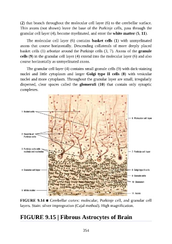
(2) that branch throughout the molecular cell layer (6) to the cerebellar surface.

               Thin axons (not shown) leave the base of the Purkinje cells, pass through the
               granular cell layer (4), become myelinated, and enter the white matter (5, 11).

                   The  molecular  cell  layer  (6)  contains  basket  cells  (1)  with  unmyelinated
               axons  that  course  horizontally.  Descending  collaterals  of  more  deeply  placed
               basket cells (1) arborize around the Purkinje cells (3, 7). Axons of the granule

               cells (9) in the granular cell layer (4) extend into the molecular layer (6) and also
               course horizontally as unmyelinated axons.

                   The granular cell layer (4) contains small granule cells (9) with dark-staining
               nuclei  and  little  cytoplasm  and  larger  Golgi  type  II  cells  (8)  with  vesicular

               nuclei and more cytoplasm. Throughout the granular layer are small, irregularly
               dispersed,  clear  spaces  called  the  glomeruli  (10)  that  contain  only  synaptic
               complexes.


               FIGURE 9.14 ■ Cerebellar cortex: molecular, Purkinje cell, and granular cell
               layers. Stain: silver impregnation (Cajal method). High magnification.


               FIGURE 9.15 | Fibrous Astrocytes of Brain


                                                          354