
Page 530 - Atlas of Histology with Functional Correlations
P. 530
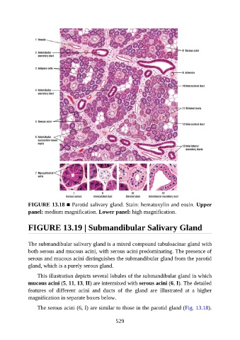
FIGURE 13.18 ■ Parotid salivary gland. Stain: hematoxylin and eosin. Upper
               panel: medium magnification. Lower panel: high magnification.


               FIGURE 13.19 | Submandibular Salivary Gland


               The submandibular salivary gland is a mixed compound tubuloacinar gland with
               both serous and mucous acini, with serous acini predominating. The presence of
               serous and mucous acini distinguishes the submandibular gland from the parotid

               gland, which is a purely serous gland.

                   This illustration depicts several lobules of the submandibular gland in which
               mucous acini (5, 11, 13, II) are intermixed with serous acini (6, I). The detailed
               features  of  different  acini  and  ducts  of  the  gland  are  illustrated  at  a  higher

               magnification in separate boxes below.

                   The serous acini (6, I) are similar to those in the parotid gland (Fig. 13.18).


                                                          529