
Page 532 - Atlas of Histology with Functional Correlations
P. 532
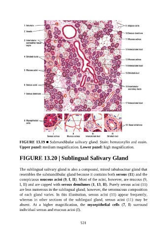
FIGURE 13.19 ■ Submandibular salivary gland. Stain: hematoxylin and eosin.
               Upper panel: medium magnification. Lower panel: high magnification.


               FIGURE 13.20 | Sublingual Salivary Gland


               The sublingual salivary gland is also a compound, mixed tubuloacinar gland that
               resembles the submandibular gland because it contains both serous (11) and the
               conspicuous mucous acini (9, I, II). Most of the acini, however, are mucous (9,

               I, II) and are capped with serous demilunes (1, 13, II). Purely serous acini (11)
               are less numerous in the sublingual gland; however, the seromucous composition
               of  each  gland  varies.  In  this  illustration,  serous  acini  (11)  appear  frequently,
               whereas  in  other  sections  of  the  sublingual  gland,  serous  acini  (11)  may  be

               absent.  At  a  higher  magnification,  the  myoepithelial  cells  (7,  I)  surround
               individual serous and mucous acini (I).


                                                          531