
Page 534 - Atlas of Histology with Functional Correlations
P. 534
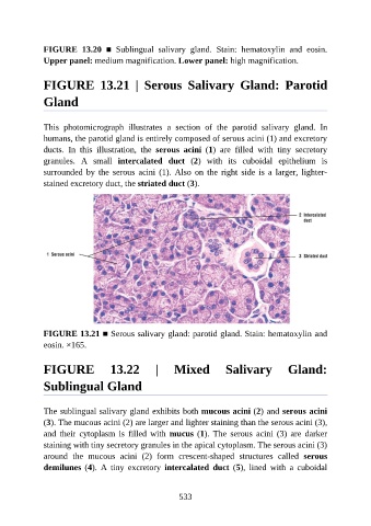
FIGURE  13.20  ■  Sublingual  salivary  gland.  Stain:  hematoxylin  and  eosin.

               Upper panel: medium magnification. Lower panel: high magnification.


               FIGURE  13.21  |  Serous  Salivary  Gland:  Parotid


               Gland


               This  photomicrograph  illustrates  a  section  of  the  parotid  salivary  gland.  In
               humans, the parotid gland is entirely composed of serous acini (1) and excretory
               ducts.  In  this  illustration,  the  serous  acini  (1)  are  filled  with  tiny  secretory

               granules.  A  small  intercalated  duct  (2)  with  its  cuboidal  epithelium  is
               surrounded  by  the  serous  acini  (1).  Also  on  the  right  side  is  a  larger,  lighter-
               stained excretory duct, the striated duct (3).


               FIGURE 13.21 ■ Serous salivary gland: parotid gland. Stain: hematoxylin and
               eosin. ×165.


               FIGURE  13.22  |  Mixed  Salivary  Gland:

               Sublingual Gland


               The sublingual salivary gland exhibits both mucous acini (2) and serous acini
               (3). The mucous acini (2) are larger and lighter staining than the serous acini (3),
               and  their  cytoplasm  is  filled  with  mucus (1).  The  serous  acini  (3)  are  darker
               staining with tiny secretory granules in the apical cytoplasm. The serous acini (3)

               around  the  mucous  acini  (2)  form  crescent-shaped  structures  called  serous
               demilunes (4).  A  tiny  excretory  intercalated  duct  (5),  lined  with  a  cuboidal


                                                          533