
Page 535 - Atlas of Histology with Functional Correlations
P. 535
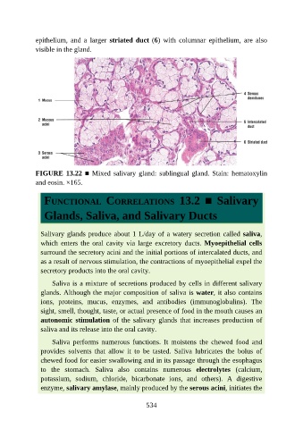
epithelium,  and  a  larger  striated duct (6)  with  columnar  epithelium,  are  also

               visible in the gland.


               FIGURE 13.22 ■ Mixed salivary gland: sublingual gland. Stain: hematoxylin
               and eosin. ×165.


                  FUNCTIONAL  CORRELATIONS  13.2  ■  Salivary


                  Glands, Saliva, and Salivary Ducts


                 Salivary glands produce about 1 L/day of a watery secretion called saliva,
                 which enters the oral cavity via large excretory ducts. Myoepithelial cells
                 surround the secretory acini and the initial portions of intercalated ducts, and

                 as a result of nervous stimulation, the contractions of myoepithelial expel the
                 secretory products into the oral cavity.

                     Saliva is a mixture of secretions produced by cells in different salivary
                 glands. Although the major composition of saliva is water, it also contains
                 ions,  proteins,  mucus,  enzymes,  and  antibodies  (immunoglobulins).  The

                 sight, smell, thought, taste, or actual presence of food in the mouth causes an
                 autonomic stimulation  of  the  salivary  glands  that  increases  production  of
                 saliva and its release into the oral cavity.

                     Saliva  performs  numerous  functions.  It  moistens  the  chewed  food  and

                 provides  solvents  that  allow  it  to  be  tasted.  Saliva  lubricates  the  bolus  of
                 chewed food for easier swallowing and in its passage through the esophagus
                 to  the  stomach.  Saliva  also  contains  numerous  electrolytes  (calcium,
                 potassium,  sodium,  chloride,  bicarbonate  ions,  and  others).  A  digestive
                 enzyme, salivary amylase, mainly produced by the serous acini, initiates the


                                                          534