
Page 61 - DP Vol 20 No 5_Neat
P. 61
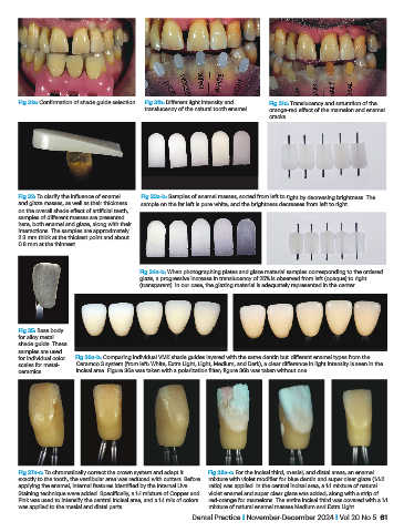
Fig 31a: Confirmation of shade guide selection.  Fig 31b: Different light intensity and   Fig 31c: Translucency and saturation of the
                                                translucency of the natural tooth enamel.  orange-red effect of the mamelon and enamel
                                                                                     cracks.


           Fig 32: To clarify the influence of enamel   Fig 33a-b: Samples of enamel masses, sorted from left to right by decreasing brightness. The
           and glaze masses, as well as their thickness   sample on the far left is pure white, and the brightness decreases from left to right.
           on the overall shade effect of artificial teeth,
           samples of different masses are presented
           here, both enamel and glaze, along with their
           interactions. The samples are approximately
           2.3 mm thick at the thickest point and about
           0.8 mm at the thinnest.


                                               Fig 34a-b: When photographing plates and glaze material samples corresponding to the ordered
                                               glaze, a progressive increase in translucency of 25% is observed from left (opaque) to right
                                               (transparent). In our case, the glazing material is adequately represented in the center.


           Fig 35: Base body
           for alloy metal
           shade guide. These
           samples are used
           for individual color   Fig 36a-b: Comparing individual VMK shade guides layered with the same dentin but different enamel types from the
           scales for metal-  Ceramco 3 system (from left: White, Extra Light, Light, Medium, and Dark), a clear difference in light intensity is seen in the
           ceramics.        incisal area. Figure 36a was taken with a polarization filter; figure 36b was taken without one.


           Fig 37a-c: To chromatically correct the crown system and adapt it   Fig 38a-c: For the incisal third, mesial, and distal areas, an enamel
           exactly to the tooth, the vestibular area was reduced with cutters. Before   mixture with violet modifier for blue dentin and super clear glaze (1:1:2
           applying the enamel, internal features identified by the Internal Live   ratio) was applied. In the central incisal area, a 1:1 mixture of natural
           Staining technique were added. Specifically, a 1:1 mixture of Copper and   violet enamel and super clear glaze was added, along with a strip of
           Pink was used to intensify the central incisal area, and a 1:1 mix of colors   red-orange for mamelons. The entire incisal third was covered with a 1:1
           was applied to the mesial and distal parts.             mixture of natural enamel masses Medium and Extra Light.
                                                              Dental Practice I November-December 2024 I Vol 20 No 5  61