
Page 577 - Atlas of Small Animal CT and MRI
P. 577
Adrenal Gland 567
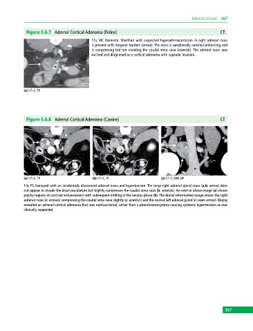
Figure 5.6.7 Adrenal Cortical Adenoma (Feline) CT
11y MC Domestic Shorthair with suspected hyperadrenocorticism. A right adrenal mass
is present with irregular borders (arrow). The mass is peripherally contrast enhancing and
is compressing but not invading the caudal vena cava (asterisk). The adrenal mass was
excised and diagnosed as a cortical adenoma with capsular invasion.
(a) CT+C, TP
Figure 5.6.8 Adrenal Cortical Adenoma (Canine) CT
(a) CT+C, TP (b) CT+C, TP (c) CT+C, MIP, DP
10y FS Samoyed with an incidentally discovered adrenal mass and hypertension. The large right adrenal gland mass (a,b: arrow) does
not appear to invade the local vasculature but slightly compresses the caudal vena cava (b: asterisk). An arterial phase image (a) shows
patchy regions of contrast enhancement with subsequent infilling in the venous phase (b). The dorsal reformatted image shows the right
adrenal mass (c: arrows) compressing the caudal vena cava slightly (c: asterisk) and the normal left adrenal gland (c: open arrow). Biopsy
revealed an adrenal cortical adenoma that was nonfunctional, rather than a pheochromocytoma causing systemic hypertension as was
clinically suspected.
567