
Page 576 - Atlas of Small Animal CT and MRI
P. 576
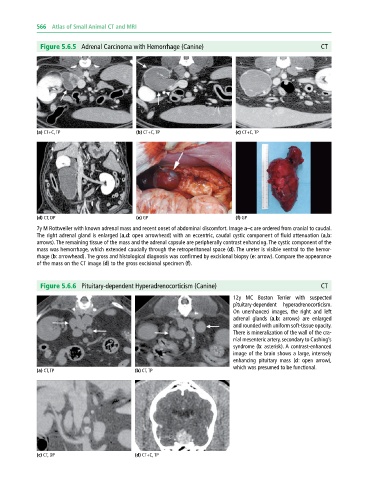
566  Atlas of Small Animal CT and MRI


              Figure 5.6.5  Adrenal Carcinoma with Hemorrhage (Canine)                                     CT


             (a) CT+C, TP                     (b) CT+C, TP                     (c) CT+C, TP


             (d) CT, DP                       (e) GP                           (f) GP
             7y M Rottweiler with known adrenal mass and recent onset of abdominal discomfort. Image a–c are ordered from cranial to caudal.
             The right adrenal gland is enlarged (a,d: open arrowhead) with an eccentric, caudal cystic component of fluid attenuation (a,b:
             arrows). The remaining tissue of the mass and the adrenal capsule are peripherally contrast enhancing. The cystic component of the
             mass was hemorrhage, which extended caudally through the retroperitoneal space (d). The ureter is visible ventral to the hemor-
             rhage (b: arrowhead). The gross and histological diagnosis was confirmed by excisional biopsy (e: arrow). Compare the appearance
             of the mass on the CT image (d) to the gross excisional specimen (f).


              Figure 5.6.6  Pituitary-dependent Hyperadrenocorticism (Canine)                              CT

                                                                              12y MC Boston  Terrier with suspected
                                                                                pituitary‐dependent  hyperadrenocorticism.
                                                                              On unenhanced images, the right and left
                                                                              adrenal glands (a,b: arrows) are enlarged
                                                                              and rounded with uniform soft‐tissue opacity.
                                                                              There is mineralization of the wall of the cra-
                                                                              nial mesenteric artery, secondary to Cushing’s
                                                                              syndrome (b: asterisk). A contrast‐enhanced
                                                                              image of the brain shows a large, intensely
                                                                              enhancing pituitary mass (d: open arrow),
                                                                              which was presumed to be functional.
             (a) CT, TP                       (b) CT, TP


             (c) CT, DP                       (d) CT+C, TP