
Page 580 - Atlas of Small Animal CT and MRI
P. 580
570 Atlas of Small Animal CT and MRI
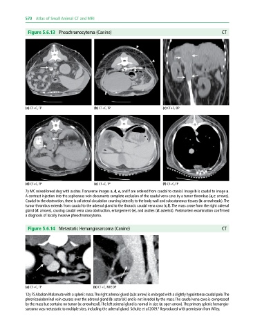
Figure 5.6.13 Pheochromocytoma (Canine) CT
(a) CT+C, TP (b) CT+C, TP (c) CT+C, DP
(d) CT+C, TP (e) CT+C, TP (f) CT+C, TP
7y MC mixed‐breed dog with ascites. Transverse images a, d, e, and f are ordered from caudal to cranial. Image b is caudal to image a.
A contrast injection into the saphenous vein documents complete occlusion of the caudal vena cava by a tumor thrombus (a,c: arrows).
Caudal to the obstruction, there is collateral circulation coursing laterally to the body wall and subcutaneous tissues (b: arrowheads). The
tumor thrombus extends from caudal to the adrenal gland to the thoracic caudal vena cava (c,f). The mass arose from the right adrenal
gland (d: arrows), causing caudal vena cava obstruction, enlargement (e), and ascites (d: asterisk). Postmortem examination confirmed
a diagnosis of locally invasive pheochromocytoma.
Figure 5.6.14 Metastatic Hemangiosarcoma (Canine) CT
(a) CT+C, TP (b) CT+C, MIP, DP
12y FS Alaskan Malamute with a splenic mass. The right adrenal gland (a,b: arrow) is enlarged with a slightly hypointense caudal pole. The
phrenicoabdominal vein courses over the adrenal gland (b: asterisk) and is not invaded by the mass. The caudal vena cava is compressed
by the mass but contains no tumor (a: arrowhead). The left adrenal gland is normal in size (a: open arrow). The primary splenic hemangio-
8
sarcoma was metastatic to multiple sites, including the adrenal gland. Schultz et al 2009. Reproduced with permission from Wiley.