
Page 584 - Atlas of Small Animal CT and MRI
P. 584
574 Atlas of Small Animal CT and MRI
are typically uniformly and moderately contrast enhanc values of greater than 55 are more likely with benign
ing and have variable margin definition (Figure 5.7.16). nodules.
Nodules are generally less than 1–2 cm in diameter, and On MR images, benign splenic nodules, including
multiple nodules may be distributed throughout the lymphoid hyperplasia and extramedullary hemat
splenic parenchyma. Larger masses may also occur in opoiesis, are hypointense on T1 and T2 images, with
some cases (Figure 5.7.17). decreased enhancement relative to normal splenic
Lymphoid hyperplasia arises from the splenic white parenchyma. 11
pulp and may be up to 5–6 cm in diameter. Larger, mass‐ Mineralization of the spleen may occur in small foci
like nodules can distort the splenic capsule and may or in a lacy pattern secondary to hyperadrenocorticism
have a nonuniform, stellate contrast‐enhancement pat or chronic steroid administration (Figure 5.7.20). This is
tern (Figures 5.7.18, 5.7.19). Contrast‐enhanced HU a benign finding.
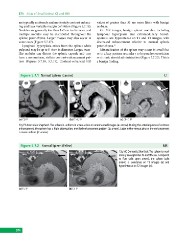
Figure 5.7.1 Normal Spleen (Canine) CT
(a) CT, TP (b) CT+C, TP (c) CT+C, TP
10y FS Australian Shepherd. The spleen is uniform in attenuation on unenhanced images (a: arrow). During the arterial phase of contrast
enhancement, the spleen has a high‐attenuation, mottled enhancement pattern (b: arrow). Later in the venous phase, the enhancement
is more uniform (c: arrow).
Figure 5.7.2 Normal Spleen (Feline) MR
12y MC Domestic Shorthair. The spleen is mod
erately enlarged due to anesthesia. Compared
to liver (a,b: open arrow), the spleen (a,b:
arrow) is isointense on T1 images (a) and
hyperintense on T2 images (b).
(a) T1, TP (b) T2, TP
574