
Page 587 - Atlas of Small Animal CT and MRI
P. 587
Spleen 577
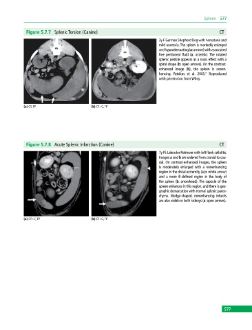
Figure 5.7.7 Splenic Torsion (Canine) CT
3y F German Shepherd Dog with hematuria and
mild anorexia. The spleen is markedly enlarged
and hypoattenuating (a: arrows) with associated
free peritoneal fluid (a: asterisk). The rotated
splenic pedicle appears as a mass effect with a
spiral shape (b: open arrows). On the contrast‐
enhanced image (b), the spleen is nonen
hancing. Patsikas et al. 2005. Reproduced
5
with permission from Wiley.
(a) CT, TP (b) CT+C, TP
Figure 5.7.8 Acute Splenic Infarction (Canine) CT
7y FS Labrador Retriever with left flank cellulitis.
Images a and b are ordered from cranial to cau
dal. On contrast‐enhanced images, the spleen
is moderately enlarged with a nonenhancing
region in the distal extremity (a,b: white arrow)
and a more ill‐defined region in the body of
the spleen (b: arrowhead). The capsule of the
spleen enhances in this region, and there is geo
graphic demarcation with normal splenic paren
chyma. Wedge‐shaped, nonenhancing infarcts
are also visible in both kidneys (a: open arrows).
(a) CT+C, TP (b) CT+C, TP
577