
Page 590 - Atlas of Small Animal CT and MRI
P. 590
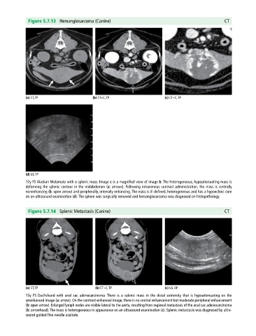
Figure 5.7.13 Hemangiosarcoma (Canine) CT
(a) CT, TP (b) CT+C, TP (c) CT+C, TP
(d) US, TP
12y FS Alaskan Malamute with a splenic mass. Image c is a magnified view of image b. The heterogeneous, hypoattenuating mass is
deforming the splenic contour in the midabdomen (a: arrows). Following intravenous contrast administration, the mass is centrally
nonenhancing (b: open arrow) and peripherally, intensely enhancing. The mass is ill defined, heterogeneous and has a hypoechoic core
on an ultrasound examination (d). The spleen was surgically removed and hemangiosarcoma was diagnosed on histopathology.
Figure 5.7.14 Splenic Metastasis (Canine) CT
(a) CT, TP (b) CT+C, TP (c) US, OP
15y FS Dachshund with anal sac adenocarcinoma. There is a splenic mass in the distal extremity that is hypoattenuating on the
unenhanced image (a: arrow). On the contrast‐enhanced image, there is no central enhancement but moderate peripheral enhancement
(b: open arrow). Enlarged lymph nodes are visible lateral to the aorta, resulting from regional metastasis of the anal sac adenocarcinoma
(b: arrowhead). The mass is heterogeneous in appearance on an ultrasound examination (c). Splenic metastasis was diagnosed by ultra
sound‐guided fine‐needle aspirate.