
Page 585 - Atlas of Small Animal CT and MRI
P. 585
Spleen 575
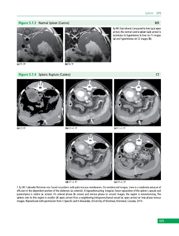
Figure 5.7.3 Normal Spleen (Canine) MR
8y MC Dachshund. Compared to liver (a,b: open
arrow), the normal canine spleen (a,b: arrow) is
isointense to hypointense to liver on T1 images
(a) and hyperintense on T2 images (b).
(a) T1, TP (b) T2, TP
Figure 5.7.4 Splenic Rupture (Canine) CT
(a) CT, TP (b) CT+C, TP (c) CT+C, TP
(d) CT+C, TP (e) CT+C, TP
1.5y MC Labrador Retriever mix found recumbent with pale mucous membranes. On unenhanced images, there is a moderate amount of
effusion in the dependent portion of the abdomen (a: asterisk). A hypoattenuating, irregular, linear separation of the splenic capsule and
parenchyma is visible (a: arrow). On arterial phase (b: arrow) and venous phase (c: arrow) images, the region is nonenhancing. The
splenic vein to this region is smaller (d: open arrow) than a neighboring intraparenchymal vessel (e: open arrow) on late‐phase venous
images. Reproduced with permission from S Specchi and K Alexander, University of Montreal, Montreal, Canada, 2014.
575