
Page 589 - Atlas of Small Animal CT and MRI
P. 589
Spleen 579
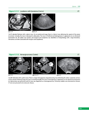
Figure 5.7.11 Lymphoma with Hematoma (Canine) CT
(a) CT, TP (b) US, SP
12y FS Labrador Retriever with a splenic mass. On an unenhanced image, there is a lobular mass deforming the capsule of the spleen
(a: arrows). Centrally, the mass is decreased in attenuation. The mass is ill defined and heterogeneous in appearance on an ultrasound
examination (b). The spleen was excised, and marginal zone lymphoma was identified on histopathology with a large hematoma.
Hematomas are often associated with marginal zone lymphomas.
Figure 5.7.12 Hemangiosarcoma (Canine) CT
(a) CT, TP (b) CT+C, TP (c) US, SP
13y MC Dalmatian with a splenic mass. There is a large, heterogeneous, hypoattenuating mass deforming the splenic capsule (a: arrows).
On the contrast‐enhanced image, the mass is nonenhancing (b). The mass is heterogeneous in appearance on an ultrasound examination
(c). Splenectomy was performed, and the mass was diagnosed as a hemangiosarcoma. Pulmonary nodules were discovered on thoracic
CT and were presumed to be metastatic disease.
579