Page 591 - Atlas of Small Animal CT and MRI
P. 591

Spleen  581


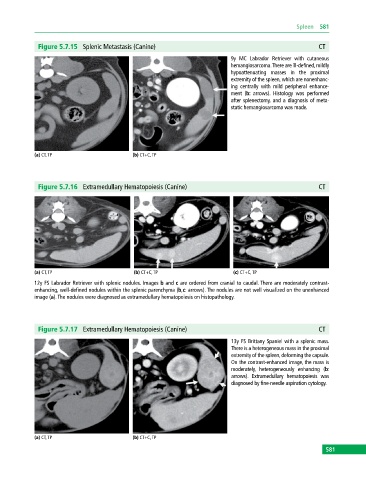
              Figure 5.7.15  Splenic Metastasis (Canine)                                                  CT
                                                                             9y MC Labrador Retriever with cutaneous
                                                                             hemangiosarcoma. There are ill‐defined, mildly
                                                                             hypoattenuating masses in the proximal
                                                                             extremity of the spleen, which are nonenhanc­
                                                                             ing centrally with mild peripheral enhance­
                                                                             ment (b: arrows). Histology was  performed
                                                                             after splenectomy, and a diagnosis of meta­
                                                                             static hemangiosarcoma was made.


            (a) CT, TP                       (b) CT+C, TP


              Figure 5.7.16  Extramedullary Hematopoiesis (Canine)                                        CT


            (a) CT, TP                       (b) CT+C, TP                     (c) CT+C, TP
            12y FS Labrador Retriever with splenic nodules. Images b and c are ordered from cranial to caudal. There are moderately contrast‐
            enhancing, well‐defined nodules within the splenic parenchyma (b,c: arrows). The nodules are not well visualized on the unenhanced
            image (a). The nodules were diagnosed as extramedullary hematopoiesis on histopathology.


              Figure 5.7.17  Extramedullary Hematopoiesis (Canine)                                        CT

                                                                             13y FS Brittany Spaniel with a splenic mass.
                                                                             There is a heterogeneous mass in the proximal
                                                                             extremity of the spleen, deforming the capsule.
                                                                             On the contrast‐enhanced image, the mass is
                                                                             moderately, heterogeneously enhancing (b:
                                                                             arrows). Extramedullary hematopoiesis was
                                                                             diagnosed by fine‐needle aspiration cytology.


            (a) CT, TP                       (b) CT+C, TP
                                                                                                             581
   586   587   588   589   590   591   592   593   594   595   596