
Page 592 - Atlas of Small Animal CT and MRI
P. 592
582 Atlas of Small Animal CT and MRI
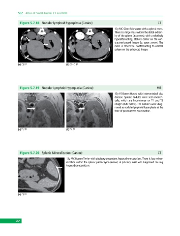
Figure 5.7.18 Nodular lymphoid hyperplasia (Canine) CT
13y MC Giant Schnauzer with a splenic mass.
There is a large mass within the distal extrem
ity of the spleen (a: arrows), with a relatively
hypoattenuating, stellate center on the con
trast‐enhanced image (b: open arrow). The
mass is otherwise isoattenuating to normal
spleen on the enhanced image.
(a) CT, TP (b) CT+C, TP
Figure 5.7.19 Nodular Lymphoid Hyperplasia (Canine) MR
13y FS Basset Hound with intervertebral disc
disease. Splenic nodules were seen inciden
tally, which are hypointense on T1 and T2
images (a,b: arrow). The nodules were diag
nosed as nodular lymphoid hyperplasia at the
time of postmortem examination.
(a) T1, TP (b) T2, TP
Figure 5.7.20 Splenic Mineralization (Canine) CT
12y MC Boston Terrier with pituitary‐dependent hyperadrenocorticism. There is lacy miner
alization within the splenic parenchyma (arrow). A pituitary mass was diagnosed causing
hyperadrenocorticism.
(a) CT, TP
582