
Page 579 - Atlas of Small Animal CT and MRI
P. 579
Adrenal Gland 569
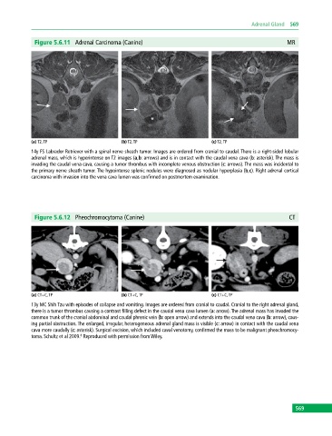
Figure 5.6.11 Adrenal Carcinoma (Canine) MR
(a) T2, TP (b) T2, TP (c) T2, TP
14y FS Labrador Retriever with a spinal nerve sheath tumor. Images are ordered from cranial to caudal. There is a right‐sided lobular
adrenal mass, which is hyperintense on T2 images (a,b: arrows) and is in contact with the caudal vena cava (b: asterisk). The mass is
invading the caudal vena cava, causing a tumor thrombus with incomplete venous obstruction (c: arrows). The mass was incidental to
the primary nerve sheath tumor. The hypointense splenic nodules were diagnosed as nodular hyperplasia (b,c). Right adrenal cortical
carcinoma with invasion into the vena cava lumen was confirmed on postmortem examination.
Figure 5.6.12 Pheochromocytoma (Canine) CT
(a) CT+C, TP (b) CT+C, TP (c) CT+C, TP
13y MC Shih Tzu with episodes of collapse and vomiting. Images are ordered from cranial to caudal. Cranial to the right adrenal gland,
there is a tumor thrombus causing a contrast filling defect in the caudal vena cava lumen (a: arrow). The adrenal mass has invaded the
common trunk of the cranial abdominal and caudal phrenic vein (b: open arrow) and extends into the caudal vena cava (b: arrow), caus-
ing partial obstruction. The enlarged, irregular, heterogeneous adrenal gland mass is visible (c: arrow) in contact with the caudal vena
cava more caudally (c: asterisk). Surgical excision, which included caval venotomy, confirmed the mass to be malignant pheochromocy-
toma. Schultz et al 2009. Reproduced with permission from Wiley.
8
569