
Page 574 - Atlas of Small Animal CT and MRI
P. 574
564 Atlas of Small Animal CT and MRI
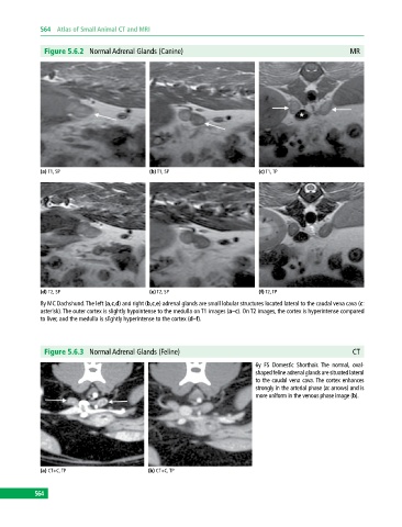
Figure 5.6.2 Normal Adrenal Glands (Canine) MR
(a) T1, SP (b) T1, SP (c) T1, TP
(d) T2, SP (e) T2, SP (f) T2, TP
8y MC Dachshund. The left (a,c,d) and right (b,c,e) adrenal glands are small lobular structures located lateral to the caudal vena cava (c:
asterisk). The outer cortex is slightly hypointense to the medulla on T1 images (a–c). On T2 images, the cortex is hyperintense compared
to liver, and the medulla is slightly hyperintense to the cortex (d–f).
Figure 5.6.3 Normal Adrenal Glands (Feline) CT
6y FS Domestic Shorthair. The normal, oval‐
shaped feline adrenal glands are situated lateral
to the caudal vena cava. The cortex enhances
strongly in the arterial phase (a: arrows) and is
more uniform in the venous phase image (b).
(a) CT+C, TP (b) CT+C, TP
564