
Page 570 - Atlas of Small Animal CT and MRI
P. 570
560 Atlas of Small Animal CT and MRI
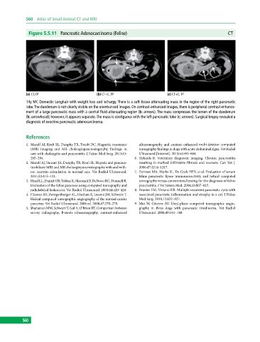
Figure 5.5.11 Pancreatic Adenocarcinoma (Feline) CT
(a) CT, TP (b) CT+C, TP (c) CT+C, TP
14y MC Domestic Longhair with weight loss and lethargy. There is a soft‐tissue attenuating mass in the region of the right pancreatic
lobe. The duodenum is not clearly visible on the unenhanced images. On contrast‐enhanced images, there is peripheral contrast enhance-
ment of a large pancreatic mass with a central fluid‐attenuating region (b: arrows). The mass compresses the lumen of the duodenum
(b: arrowhead); however, it appears separate. The mass is contiguous with the left pancreatic lobe (c: arrows). Surgical biopsy revealed a
diagnosis of exocrine pancreatic adenocarcinoma.
References
1. Marolf AJ, Kraft SL, Dunphy TR, Twedt DC. Magnetic resonance ultrasonography and contrast‐enhanced multi‐detector computed
(MR) imaging and MR cholangiopancreatography findings in tomography findings in dogs with acute abdominal signs. Vet Radiol
cats with cholangitis and pancreatitis. J Feline Med Surg. 2013;15: Ultrasound [Internet]. 2013;54:591–604.
285–294. 6. Hylands R. Veterinary diagnostic imaging. Chronic pancreatitis
2. Marolf AJ, Stewart JA, Dunphy TR, Kraft SL. Hepatic and pancrea resulting in marked infiltrative fibrosis and necrosis. Can Vet J.
ticobiliary MRI and MR cholangiopancreatography with and with 2006;47:1214–1217.
out secretin stimulation in normal cats. Vet Radiol Ultrasound. 7. Forman MA, Marks SL, De Cock HEV, et al. Evaluation of serum
2011;52:415–421. feline pancreatic lipase immunoreactivity and helical computed
3. Head LL, Daniel GB, Tobias K, Morandi F, DeNovo RC, Donnell R. tomography versus conventional testing for the diagnosis of feline
Evaluation of the feline pancreas using computed tomography and pancreatitis. J Vet Intern Med. 2004;18:807–815.
radiolabeled leukocytes. Vet Radiol Ultrasound. 2003;44:420–428. 8. Branter EM, Viviano KR. Multiple recurrent pancreatic cysts with
4. Cáceres AV, Zwingenberger AL, Hardam E, Lucena JM, Schwarz T. associated pancreatic inflammation and atrophy in a cat. J Feline
Helical computed tomographic angiography of the normal canine Med Surg. 2010;12:822–827.
pancreas. Vet Radiol Ultrasound. 2006 ed. 2006;47:270–278. 9. Mai W, Cáceres AV. Dual‐phase computed tomographic angio
5. Shanaman MM, Schwarz T, Gal A, O’Brien RT. Comparison between graphy in three dogs with pancreatic insulinoma. Vet Radiol
survey radiography, B‐mode ultrasonography, contrast‐enhanced Ultrasound. 2008;49:141–148.
560