
Page 573 - Atlas of Small Animal CT and MRI
P. 573
Adrenal Gland 563
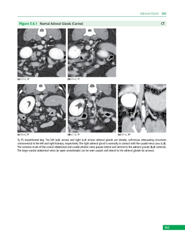
Figure 5.6.1 Normal Adrenal Glands (Canine) CT
(a) CT+C, TP (b) CT+C, TP
(c) CT+C, TP (d) CT+C, TP (e) CT+C, DP
3y FS mixed‐breed dog. The left (a,b: arrow) and right (c,d: arrow) adrenal glands are slender, soft‐tissue attenuating structures
craniomedial to the left and right kidneys, respectively. The right adrenal gland is normally in contact with the caudal vena cava (c,d).
The common trunk of the cranial abdominal and caudal phrenic veins passes lateral and ventral to the adrenal glands (b,d: asterisk).
The large cranial abdominal veins (e: open arrowheads) can be seen caudal and lateral to the adrenal glands (e: arrows).
563