
Page 568 - Atlas of Small Animal CT and MRI
P. 568
558 Atlas of Small Animal CT and MRI
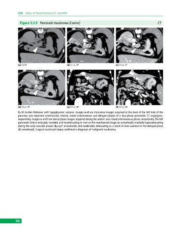
Figure 5.5.9 Pancreatic Insulinoma (Canine) CT
(a) CT, TP (b) CT+C, TP (c) CT+C, TP
(d) CT+C, TP (e) CT+C, DP (f) CT+C, DP
8y M Golden Retriever with hypoglycemic seizures. Images a–d are transverse images acquired at the level of the left limb of the
pancreas and represent unenhanced, arterial, mixed arteriovenous and delayed phases of a four‐phase pancreatic CT angiogram,
respectively. Images e and f are dorsal plane images acquired during the arterial and mixed arteriovenous phase, respectively. The left
pancreatic limb is enlarged, rounded, and isoattenuating to liver on the unenhanced image (a: arrowhead); markedly hyperatenuating
during the early vascular phases (b,c,e,f: arrowhead); and moderately attenuating as a result of slow washout in the delayed phase
(d: arrowhead). Surgical excisional biopsy confirmed a diagnosis of malignant insulinoma.
558