
Page 569 - Atlas of Small Animal CT and MRI
P. 569
Pancreas 559
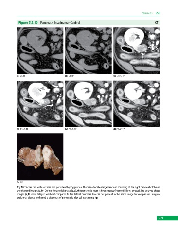
Figure 5.5.10 Pancreatic Insulinoma (Canine) CT
(a) CT, TP (b) CT, TP (c) CT+C, TP
(d) CT+C, TP (e) CT+C, TP (f) CT+C, TP
(g) GP
10y MC Terrier mix with seizures and persistent hypoglycemia. There is a focal enlargement and rounding of the right pancreatic lobe on
unenhanced images (a,b). During the arterial phase (c,d), the pancreatic mass is hypoattenuating medially (c: arrows). The delayed phase
images (e,f) show delayed washout compared to the lateral pancreas. Liver is not present in the same image for comparison. Surgical
excisional biopsy confirmed a diagnosis of pancreatic islet cell carcinoma (g).
559