Page 567 - Atlas of Small Animal CT and MRI
P. 567

Pancreas  557


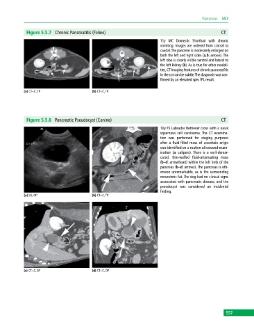
              Figure 5.5.7  Chronic Pancreatitis (Feline)                                                 CT
                                                                             11y MC Domestic Shorthair with chronic
                                                                               vomiting. Images are ordered from cranial to
                                                                             caudal. The pancreas is moderately enlarged on
                                                                             both the left and right sides (a,b: arrows). The
                                                                             left lobe is clearly visible ventral and lateral to
                                                                             the left kidney (b). As is true for other modali-
                                                                             ties, CT imaging features of chronic pancreatitis
                                                                             in the cat can be subtle. The  diagnosis was con-
                                                                             firmed by an elevated spec fPL result.

            (a) CT+C, TP                     (b) CT+C, TP


              Figure 5.5.8  Pancreatic Pseudocyst (Canine)                                                CT
                                                                             10y FS Labrador Retriever cross with a nasal
                                                                             squamous  cell carcinoma. The CT examina-
                                                                             tion was performed for staging purposes
                                                                             after a fluid‐filled mass of uncertain origin
                                                                             was identified on a routine ultrasound exam-
                                                                             ination (a: calipers). There is a well‐demar-
                                                                             cated, thin‐walled fluid‐attenuating mass
                                                                             (b–d: arrowhead) within the left limb of the
                                                                             pancreas (b–d: arrows). The pancreas is oth-
                                                                             erwise unremarkable, as is the surrounding
                                                                             mesenteric fat. The dog had no clinical signs
                                                                             associated with pancreatic disease, and the
                                                                             pseudocyst was considered an incidental
                                                                             finding.
            (a) US, OP                       (b) CT+C, TP


            (c) CT+C, SP                     (d) CT+C, DP


                                                                                                             557
   562   563   564   565   566   567   568   569   570   571   572