
Page 566 - Atlas of Small Animal CT and MRI
P. 566
556 Atlas of Small Animal CT and MRI
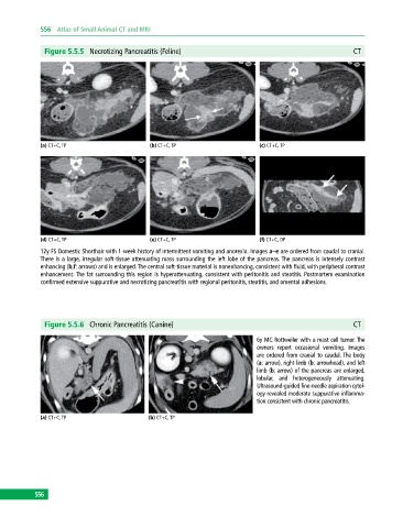
Figure 5.5.5 Necrotizing Pancreatitis (Feline) CT
(a) CT+C, TP (b) CT+C, TP (c) CT+C, TP
(d) CT+C, TP (e) CT+C, TP (f) CT+C, DP
12y FS Domestic Shorthair with 1‐week history of intermittent vomiting and anorexia. Images a–e are ordered from caudal to cranial.
There is a large, irregular soft‐tissue attenuating mass surrounding the left lobe of the pancreas. The pancreas is intensely contrast
enhancing (b,f: arrows) and is enlarged. The central soft‐tissue material is nonenhancing, consistent with fluid, with peripheral contrast
enhancement. The fat surrounding this region is hyperattenuating, consistent with peritonitis and steatitis. Postmortem examination
confirmed extensive suppurative and necrotizing pancreatitis with regional peritonitis, steatitis, and omental adhesions.
Figure 5.5.6 Chronic Pancreatitis (Canine) CT
6y MC Rottweiler with a mast cell tumor. The
owners report occasional vomiting. Images
are ordered from cranial to caudal. The body
(a: arrow), right limb (b: arrowhead), and left
limb (b: arrow) of the pancreas are enlarged,
lobular, and heterogeneously attenuating.
Ultrasound‐guided fine‐needle aspiration cytol-
ogy revealed moderate suppurative inflamma-
tion consistent with chronic pancreatitis.
(a) CT+C, TP (b) CT+C, TP
556