
Page 565 - Atlas of Small Animal CT and MRI
P. 565
Pancreas 555
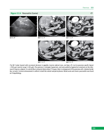
Figure 5.5.4 Pancreatitis (Canine) CT
(a) US, OP (b) CT+C, TP (c) CT+C, TP
(d) CT+C, TP (e) CT+C, TP
15y MC Cocker Spaniel with occasional decrease in appetite, invasive adrenal mass, and Spec cPL (canine pancreas‐specific lipase)
>1000 μg/L (normal range 0–200 μg/L). The pancreas is enlarged, hypoechoic, and surrounded by hyperechoic omentum on the ultra-
sound image (a: calipers). On arterial (b,c) and portal (d,e) phase CT images, there is enlargement of the pancreas with a lobular border
(b,c: arrows). Contrast enhancement is uniform in both the arterial and portal phases. Mixed acute and chronic pancreatitis was found
on histopathology.
555