
Page 38 - Rapid Review of ECG Interpretation in Small Animal Practice, 2nd Edition
P. 38
Approach to Evaluating Arrhythmias
VetBooks.ir
II
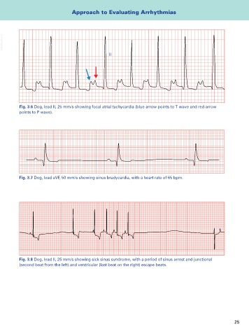
Fig. 3.6 Dog, lead II, 25 mm/s showing focal atrial tachycardia (blue arrow points to T wave and red arrow
points to P wave).
Fig. 3.7 Dog, lead aVF, 50 mm/s showing sinus bradycardia, with a heart rate of 65 bpm.
Fig. 3.8 Dog, lead II, 25 mm/s showing sick sinus syndrome, with a period of sinus arrest and junctional
(second beat from the left) and ventricular (last beat on the right) escape beats.
25