Page 575 - Veterinary Toxicology, Basic and Clinical Principles, 3rd Edition
P. 575
542 SECTION |VI Insecticids


  VetBooks.ir                                                   body tissues, MLs are largely excluded from the mamma-
                                                                  Although they have widespread distribution to most
                                                                lian brain through the action of the BBB’s p-glycoprotein
                                                                transport system (P-GP) (Fig. 43.7). The P-GP is a
                                                                170 kDa transmembrane protein coded for by the ABCB1
                                                                (formerly MDR1) gene and is located primarily in the
                                                                intestine, liver, kidney, and brain (Mosher and Court,
                                                                2010). The function of P-GP is to limit the systemic and
                                                                brain uptake of certain xenobiotics and enhance their
                                                                elimination from the body (Shen and Zhang, 2010).
                                                                Alterations in P-GP function can result in increased
                                                                systemic xenobiotic levels and enhanced penetration of
                                                                xenobiotics into the brain. MLs comprise one family of
                                                                drugs that serve as substrates for P-GP; therefore, ani-
                                                                mals with defects within the P-GP system may have
                                                                increased absorption and decreased elimination of MLs,
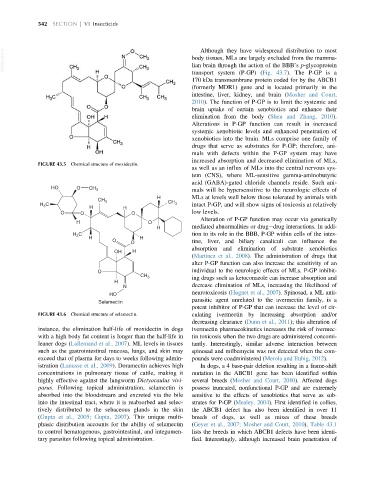
             FIGURE 43.5 Chemical structure of moxidectin.
                                                                as well as an influx of MLs into the central nervous sys-
                                                                tem (CNS), where ML-sensitive gamma-aminobutyric
                                                                acid (GABA)-gated chloride channels reside. Such ani-
                  HO      O   CH 3
                                                                mals will be hypersensitive to the neurologic effects of
                                                    H           MLs at levels well below those tolerated by animals with
                                 CH 3
              H C             H          H              CH 3    intact P-GP, and will show signs of toxicosis at relatively
               3
                     O     O                O                   low levels.
                                                                  Alteration of P-GP function may occur via genetically
                          H                      O
                                                    H           mediated abnormalities or drug drug interactions. In addi-
                         H 3 C                                  tion to its role in the BBB, P-GP within cells of the intes-
                              H                H
                                      O     O                   tine, liver, and biliary canaliculi can influence the
                                                                absorption and elimination of substrate xenobiotics
                                      OH    H
                                                                (Martinez et al., 2008). The administration of drugs that
                                                                alter P-GP function can also increase the sensitivity of an
                                 O                              individual to the neurologic effects of MLs. P-GP inhibit-
                                               CH 3             ing drugs such as ketoconazole can increase absorption and
                                      H
                                         N                      decrease elimination of MLs, increasing the likelihood of
                                                                neurotoxicosis (Hugnet et al., 2007). Spinosad, a ML anti-
                                     HO
                                                                parasitic agent unrelated to the avermectin family, is a
                                 Selamectin
                                                                potent inhibitor of P-GP that can increase the level of cir-
             FIGURE 43.6 Chemical structure of selamectin.      culating ivermectin by increasing absorption and/or
                                                                decreasing clearance (Dunn et al., 2011); this alteration of
             instance, the elimination half-life of moxidectin in dogs  ivermectin pharmacokinetics increases the risk of ivermec-
             with a high body fat content is longer than the half-life in  tin toxicosis when the two drugs are administered concomi-
             leaner dogs (Lallemand et al., 2007). ML levels in tissues  tantly. Interestingly, similar adverse interaction between
             such as the gastrointestinal mucosa, lungs, and skin may  spinosad and milbemycin was not detected when the com-
             exceed that of plasma for days to weeks following admin-  pounds were coadministered (Merola and Eubig, 2012).
             istration (Lanusse et al., 2009). Doramectin achieves high  In dogs, a 4 base-pair deletion resulting in a frame-shift
             concentrations in pulmonary tissue of cattle, making it  mutation in the ABCB1 gene has been identified within
             highly effective against the lungworm Dictyocaulus vivi-  several breeds (Mosher and Court, 2010). Affected dogs
             parus. Following topical administration, selamectin is  possess truncated, nonfunctional P-GP and are extremely
             absorbed into the bloodstream and excreted via the bile  sensitive to the effects of xenobiotics that serve as sub-
             into the intestinal tract, where it is reabsorbed and selec-  strates for P-GP (Mealey, 2004). First identified in collies,
             tively distributed to the sebaceous glands in the skin  the ABCB1 defect has also been identified in over 11
             (Gupta et al., 2005; Gupta, 2007). This unique multi-  breeds of dogs, as well as mixes of these breeds
             phasic distribution accounts for the ability of selamectin  (Geyer et al., 2007; Mosher and Court, 2010). Table 43.1
             to control hematogenous, gastrointestinal, and integumen-  lists the breeds in which ABCB1 defects have been identi-
             tary parasites following topical administration.   fied. Interestingly, although increased brain penetration of

