Page 171 - Withrow and MacEwen's Small Animal Clinical Oncology, 6th Edition
P. 171
150   PART II    Diagnostic Procedures for the Cancer Patient


  VetBooks.ir  V1     V2     V3      V79      V80     1  2  34   5  6    J1   J2     J3    J4    J5


                               V region                D region                    J region


                                  V1    V2        V3  4  J2     J3    J4    J5
            A


                             V3    4    J2                                    V4    2       J3

                             V4    2       J3                                 V4    2       J3

                            V79       4       J5                              V4    2       J3


                     40  45  50  55  60  65  70  75  80  85  90  95  100  105  110  115  120  125  130  135  140  40  45  50  55  60  65  70  75  80  85  90  95  100  105  110  115  120  125  130  135  140
              2,500                                             10,000
                                                                 9,000
              2,000                                              8,000
                                                                 7,000
              1,500                                              6,000
                                                                 5,000
              1,000                                              4,000
                                                                 3,000
                500                                              2,000
                                                                 1,000
                 0                                                  0
                              Size                                                Size
            B                                                  C
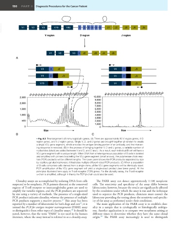
                          • Fig. 8.3  Rearrangement of immunoglobulin genes. (A) There are approximately 80 V region genes, 6 D
                          region genes, and 5 J region genes. Single V, D, and J genes are brought together at random to create
                          a single VDJ gene segment, which encodes the antigen-binding portion of an antibody, and the interven-
                          ing sequence is removed. (B) In the process of bringing together V, D and J genes, a variable number of
                          nucleotides (black) are added between V and D, and D and J. As a result, each individual B cell will have a
                          VDJ gene segment with a unique length. When DNA from a heterogeneous population of B cells is isolated
                          and amplified with primers bracketing the VDJ gene segment (small arrows), the polymerase chain reac-
                          tion (PCR) products will be different lengths. The lower panel shows the PCR products separated by size
                          by capillary gel electrophoresis; it illustrates multiple different-sized PCR products. (C) When a population
                          of B cells comprises cells derived from a single clone, all the VDJ gene segments will be identically sized.
                          PCR amplification of the VDJ gene segment will yield a single-sized product (see lower panel). All the
                          principles illustrated here apply to T-cell receptor (TCR) genes. For the clonality assay, the T-cell receptor
                          γ-chain is amplified, although in theory the TCR β-chain could also be used.
            Clonality assays are accomplished by isolating DNA from cells   The PARR assay  can detect approximately 1:100 neoplastic
         suspected to be neoplastic. PCR primers directed at the conserved   cells. The sensitivity and specificity of the assay differ between
         regions of T-cell receptor or immunoglobulin genes are used to   laboratories; however, because the results are significantly affected
         amplify the variable regions, and the PCR products are separated   by the conditions under which the assay is run and the technique
         by size using a variety of methods. The presence of a single-sized   used to separate the PCR products, clinicians must consult the
         PCR product indicates clonality, whereas the presence of multiple   laboratory providing the testing about the sensitivity and specific-
                                           27
         PCR products supports a reactive process.  This assay has been   ity of the assay as performed under their conditions.
         reported by a number of laboratories for both dogs and cats ; it is   The main application of the PARR assay is to establish clon-
                                                       27
         termed the PCR for antigen receptor rearrangements (PARR) assay   ality in  a sample that is cytologically  or histologically  ambigu-
         to distinguish it from other types of clonality assays.  It should be   ous. Another application is to compare two neoplasms arising at
                                                 28
         noted, however, that the term “PARR” is not used in the human   different times to determine whether they have the same clonal
                                                                     29
         literature, where the assay instead is referred to as a clonality assay.  origin.  The PARR assay increasingly is used to distinguish

