Page 173 - Withrow and MacEwen's Small Animal Clinical Oncology, 6th Edition
P. 173
152   PART II    Diagnostic Procedures for the Cancer Patient


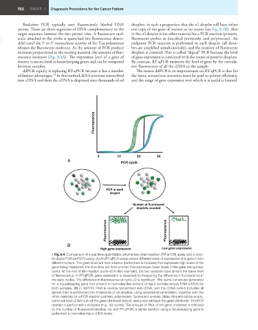
            Real-time PCR typically uses fluorescently labeled DNA   droplets, in such a proportion that the oil droplet will have either
         probes. These are short segments of DNA complementary to the   one copy of the gene of interest or no copies (see Fig. 8.4B). Also
                                                               in the oil droplet is the other material for a PCR reaction (primers,
         target sequence between the two primer sites. A fluorescent mol-
  VetBooks.ir  ecule attached to the probe is quenched (no fluorescence detect-  fluorescent probes as described previously, and polymerase). An
                                                               endpoint PCR reaction is performed in each droplet (all drop-
         able) until the 5′ to 3′ exonuclease activity of the Taq polymerase
         releases the fluorescent molecule. As the amount of PCR product   lets are amplified simultaneously), and the number of fluorescent
         increases proportional to the starting material, the amount of fluo-  droplets is counted. This is called “digital” PCR because the level
         rescence increases (Fig. 8.4A). The expression level of a gene of   of gene expression is correlated with the count of positive droplets.
         interest is normalized to housekeeping genes and can be compared   By contrast, RT-qPCR measures the level of gene by the cumula-
         between samples.                                      tive fluorescence of all the cDNA in the sample.
            ddPCR rapidly is replacing RT-qPCR because it has a number   The reason ddPCR is an improvement on RT-qPCR is that for
         of distinct advantages.  In this method, RNA is reverse transcribed   the latter, scrupulous attention must be paid to primer efficiency,
                          43
         into cDNA and then the cDNA is dispersed into thousands of oil   and the range of gene expression over which it is useful is limited.


                                              Fluorescence


                                               0         10       20       30
                                 A                        PCR cycle


                                                   PCR in each
                                                     droplet

                                                                Number of fluorescent
                                                                  droplets counted


                                                   Fluorescence                 Fluorescence


                                 B
                                                 High gene expression         Low gene expression
                          • Fig. 8.4  Comparison of a real-time quantitative polymerase chain reaction (RT-qPCR) assay and a drop-
                          let digital PCR (ddPCR) assay. (A) An RT-qPCR assay shows different levels of expression of a gene in two
                          different tumors. The green lines are from a tumor (performed in triplicate) that expresses high levels of the
                          gene being measured; the blue lines are from a tumor that expresses lower levels of the gene being mea-
                          sured. At the end of the reaction (cycle 40 in this example), the two samples have almost the same level
                          of fluorescence. In RT-qPCR, gene expression is assessed by measuring the difference in fluorescence in
                          the early cycles. The difference in fluorescence at cycle 22 is significant. The same curves are generated
                          for a housekeeping gene (not shown) to normalize the amount of input complementary DNA (cDNA) for
                          both samples. (B) In ddPCR, RNA is reverse transcribed into cDNA, and the cDNA (which includes all
                          genes) then is partitioned into thousands of oil droplets, using special instrumentation, together with the
                          other materials for a PCR reaction (primers, polymerase, fluorescent probes). Many droplets will be empty;
                          some will have cDNA but not the gene of interest (black); and some will have the gene of interest. The PCR
                          reaction is performed to endpoint (e.g., 30 cycles). The amount of RNA of the gene of interest is reflected
                          by the number of fluorescent droplets. As with RT-qPCR, a similar reaction using a housekeeping gene is
                          performed to normalize input cDNA levels.

