
Page 287 - Zoo Animal Learning and Training
P. 287
Chapter 14 Surgical Room Skills 271
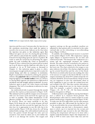
FIGURE 14.33 Left: oxygen tank yoke. Right: oxygen pressure gauge.
injection and then every 5 minutes after the injection on vaporizer settings on the gas anesthetic machine are
the anesthesia monitoring chart until the patient is adjusted for that patient and it is attached to the endo-
moved to the recovery room. Indicate on the chart when tracheal tube via the rebreathing or non‐rebreathing
the injection was given so the induction agent that hoses (Figure 14.10a-c).
follows isn’t given too soon. It is during this period that If available, the multifunctional monitor is attached
an IV catheter is placed into the cephalic, saphenous, or to the patient (Figure 14.37). The respiratory/capnog-
femoral vein depending upon the species of patient. Be raphy adapter is attached between the air hose and
ready to assist the technician by presenting the appro- endotracheal tube. This measures the respirations via a
priate leg and occluding the vessel as described in sensor and the capnograph measures the carbon
Chapter 8. Once catheterized, the patient will be hooked dioxide. The pulse oximeter is attached to the patient to
up to an IV drip set and the fluid rate will either be set measure the amount of oxygen in the blood and read
manually or via an IV fluid pump as discussed in the heart rate. The clip is attached to the tongue or any
Chapter 11. The induction agent is given to effect, place without a lot of hair. The clip has a light that picks
meaning just enough medication is injected to make the up the arterial blood flow and the color of the blood.
animal slump and offer no resistance to intubation. The flow is recorded as the heart rate reading, the color
Depth of anesthesia is checked by utilizing the patient’s is the amount of oxygen in the blood. We like to see the
reflexes. The palpebral reflex is evaluated by tapping the oxygen saturation stay about 95% and if it drops the vet-
fingertip lightly at the medial canthus of the eye. If there erinarian should be alerted immediately. Drops in heart
is a strong blink response, the patient is in a light level of rate are important to alert the veterinarian about as
anesthesia and will struggle against intubation. As more well. A guideline to use is to take the resting heart rate
induction agent is given, the reflex lessens and finally and multiply it by 35%. Take that number and subtract
disappears. The level of anesthesia is sufficient for intu- it from the resting heart rate for the lowest tolerable
bation and prep. limit. For example, a patient’s resting heart rate is
An endotracheal tube is passed through the mouth 75 bpm × .35 = 26. 75 – 26 = 49. The lowest tolerable
and into the trachea as demonstrated in Figure 14.35. limit for this patient is 49 bpm.
The assistant’s job is to pull the tongue out as far as pos- A blood pressure cuff, applied to any leg that doesn’t
sible over the incisor teeth and hold the upper jaw up have a catheter in it, measures blood pressure. The
as wide as possible. This makes visualization of the blood pressure cuffs come in a variety of sizes to accom-
pharynx possible. Once the tube is in place it needs to modate small to large companion animals (Figure 14.38).
be secured. There are many methods to do this, To make sure the fit is right either use the tape measure
Figure 14.36 shows the use of a strip of gauze roll tied provided or place the cuff around the leg and see if it
onto the tube and then around the upper jaw. Use of a can be secured within the arrows indicated on the cuff.
shoe lace tie is appropriate in case the tube has to be The monitor may also have a thermometer attach-
untied quickly. Figure 14.36 also shows the cuff being ment in which case it is inserted into the rectum and
inflated with a syringe. The cuff is inflated until you can using self‐adhesive tape secured to the tail. Otherwise,
hear the patient breath through the tube and not checking the temperature will have to be done with a
around it. Care must be taken not to over inflate the regular rectal thermometer. Expect the temperature to
cuff as it can damage the trachea. The oxygen and drop to 99°F in most cases; lower than that and the