
Page 22 - CJO_F17_GLAUCOMA_SUPPLEMENT
P. 22
C CLINICAL RESEARCH
2. Identify the width of the neuroretinal Rim
Glaucoma is defined by the loss of retinal ganglion cell axons that comprise the RNFL and neuroretinal Rim
(NRR). Diffuse or localized (particularly inferior-temporal) NRR thinning is 87% specific for glaucoma. Ex-
166
cavation or undermining of rim tissue is one of the earliest structural changes, while superior or inferior focal
notches are essentially pathognomonic for GON and predictive of rapid visual field loss that may threaten
fixation. 167-169 Focal loss is often easier to spot but is less common than diffuse loss of the neuroretinal Rim. 170,171
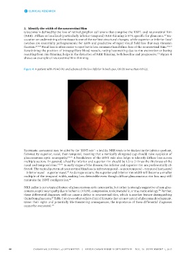
Scrutinizing the position of intrapapillary blood vessels, noting bayonetting due to rim excavation or baring
resulting from rim thinning, helps in the detection of NRR thinning, both baseline and progressive. Figure 4
172
shows an example of neuroretinal Rim thinning.
Figure 4: A patient with POAG OU and advanced rim loss inferior in both eyes, OS (b) worse than OD (a).
Systematic assessment may be aided by the ‘ISNT rule’: a healthy NRR tends to be thickest in the inferior quadrant,
followed by superior, nasal, then temporal, meaning that a vertically elongated cup should raise suspicion of
glaucomatous optic neuropathy. 45,173 A breakdown of the ISNT rule also helps to identify diffuse loss across
multiple sectors. In general, a healthy inferior and superior rim should be 1.5 to 2 times the thickness of the
nasal and temporal rims. 157,170 In early stages of the disease, the inferior and superior rim are preferentially af-
fected. The typical pattern of neuroretinal Rim loss is: inferotemporal – superotemporal – temporal horizontal
– inferior nasal – superior nasal. As damage occurs, the superior and inferior rim width will become a smaller
47
multiple of the temporal width, making loss detectable even though diffuse glaucomatous rim loss may still
maintain the ISNT configuration. 46
NRR pallor is not a typical feature of glaucomatous optic neuropathy, but rather is strongly suggestive of non-glau-
comatous optic neuropathy due to ischemic (AION), compressive, toxic/metabolic, or traumatic etiology. Further,
174
these differential diagnoses will not cause a defect in neuroretinal Rim, which is another feature distinguishing
them from glaucoma. Table 5 reviews other salient clinical features that are not typical of glaucoma development.
47
Given their sight- and potentially life-threatening consequences, the importance of these differential diagnoses
cannot be overstated.
175
22 CANADIAN JOURNAL of OPTOMETRY | REVUE CANADIENNE D’OPTOMÉTRIE VOL. 79 SUPPLEMENT 1, 2017