
Page 25 - CJO_F17_GLAUCOMA_SUPPLEMENT
P. 25
MANAGING OPEN ANGLE GLAUCOMA
4. Assess the Region of parapapillary atrophy (PPA)
There are typically 5 prominent rings that can be identified clinically on the ONH: from central to peripheral
they are the cup, the rim, the scleral Ring, zone beta and zone alpha PPA. Zone-beta parapapillary atrophy
187
(zone-β PPA) is increased scleral visibility due to degeneration of the RPE and choriocapillaris immediately
adjacent to the ONH. Zone-β PPA is rare in healthy eyes, but is more common and extensive in glaucomatous
eyes, particularly those with shallow, sloping cups. 188,189 On the contrary, zone-alpha (zone-α) PPA, irregular
pigmentary change in the RPE alone, is found in the majority of healthy eyes. When both types of PPA are
present, zone-α is always peripheral to zone-β. Zone-β PPA is larger in eyes with more advanced disease,
and spatially and temporally correlated with RNFL thinning, NRR defects, and optic disc hemorrhages. 190-193
Figure 7 illustrates the differentiation between zone-β and zone-α PPA in an eye with glaucomatous damage.
VF deterioration is more rapid in the presence of baseline zone-β PPA, and increasing PPA is associated with
progressive VF loss. The progression of PPA may be more diagnostic than its presence. 194,195 Assessing PPA
may be particularly helpful with small ONHs where intrapapillary (cupping) change is difficult to assess, and
less valuable with myopic or tilted ONHs and in older individuals where non-glaucomatous zone-β PPA may
already exist. PPA has historically been a difficult parameter to objectively quantify, but may be qualitatively
196
tracked through serial fundus photography or en face OCT images.
197
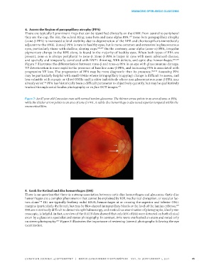
Figure 7: An 87 year-old Caucasian man with normal tension glaucoma. The thinner arrow points to an area of zone-α PPA,
while the thicker arrow points to an area of zone-β PPA. A subtle disc hemorrhage is also noted superior temporal within the
neuroretinal Rim.
5. Look for Retinal and disc hemorrhages (DH)
There is no question that there is a strong association between optic disc hemorrhages and glaucoma. Optic disc
hemorrhages are a complex phenomenon that cannot be explained by IOP, mechanical disruption, or vascular fac-
tors alone. DH are typically feathery radial RNFL hemorrhages at or crossing the superior and inferior ONH
198
margins (particularly the latter), but may be blot-shaped intrapapillary bleeds at the level of the lamina cribrosa.
199
DH are notoriously difficult to detect via ophthalmoscopy, and meticulous examination of photographs, ideally ste-
reoscopic, is helpful. In fact, a review of the OHTS data showed that only 16% of DH were detected on both clinical
exam by a glaucoma specialist and stereo photography. In contrast, 84% were overlooked on exam and noted only
on stereo photography. Figure 8 illustrates the importance of reviewing (stereo) photographs following the eye
200
examination.
CANADIAN JOURNAL of OPTOMETRY | REVUE CANADIENNE D’OPTOMÉTRIE VOL. 79 SUPPLEMENT 1, 2017 25