
Page 26 - CJO_F17_GLAUCOMA_SUPPLEMENT
P. 26
C CLINICAL RESEARCH
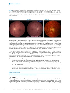
Figure 8: a) A 60 year-old woman with NTG and low ocular perfusion pressure. Recurrent disc hemorrhages were noted at
5:30 in the RNFL. Additional IOP lowering medication was added b) A 67 year-old Caucasian man with POAG. A disc hem-
orrhage was noted on the neuroretinal Rim at 6:00. IOP not at target and treatment was adjusted. c) A 76 year-old Caucasian
woman with asymmetric POAG. Recurrent DH have been noted, always OS, both at the ONH margin and at the level of the
lamina, as seen here.
Figure 8 a Figure 8 b Figure 8 c
DH are quite rare in healthy individuals (0.2 to 0.5% prevalence) but more common in those with early to moderate glau-
coma, particularly in the presence of ‘normal’ intraocular pressure. 201,202 However, given the relatively low preva-
lence of glaucoma, the majority of DH are still found in patients who have not yet been diagnosed with the disease.
It has been shown that the median time to development of a visual field defect following an optic disc hemorrhage
is 38 months. 198,203 Further, it has been suggested that more aggressive treatment after the detection of a hemorrhage
might slow down visual field progression compared to not changing treatment. Differential diagnoses include
204
venous occlusion, diabetic retinopathy, posterior vitreous detachment, ONH drusen, and AION. 205,206
DH may be the single strongest risk factor for the progression of established glaucoma and were found more com-
monly in eyes that developed glaucoma in OHTS. However, they are not considered a stand-alone diagnostic crite-
rion in the absence of other signs of glaucoma. 199,200,207-211 Despite their strong association with disease progression,
there has long been uncertainty about whether DH are a result of, or factor for progression. At present, it is gener-
ally thought that DH are a phenomenon confirming glaucoma disease activity.
Clinical Recommendations for ONH/RNFL assessment:
• Diligent and systematic clinical assessment of the ONH and RNFL is a means of early identification of
disease, and one of the cornerstones of effective glaucoma management. Particular attention should be
paid to neuroretinal Rim and RNFL changes at the superior and inferior poles, and to the identification of
optic disc hemorrhages.
• ‘We argue that ophthalmoscopy and photography remain the gold standard of imaging due to portability, ease of
interpretation, and the presence of a large database of images for comparison.’ (Spaeth GL, Reddy SC; 2014).
ANCILLARY TESTING
SPECTRAL DOMAIN OPTICAL COHERENCE TOMOGRAPHY
RNFL and ONH
Clinical (subjective) assessment of the optic nerve head (ONH) and Retinal nerve fiber layer (RNFL) is critical but
challenging: even among glaucoma specialists, significant inter- and intra-observer variability is the rule rather than
the exception. Ancillary objective imaging (most commonly optical coherence tomography, OCT) has become an
212
invaluable complement in the diagnosis of glaucoma, detecting structural change up to six years before visual field
26 CANADIAN JOURNAL of OPTOMETRY | REVUE CANADIENNE D’OPTOMÉTRIE VOL. 79 SUPPLEMENT 1, 2017