
Page 23 - CJO_F17_GLAUCOMA_SUPPLEMENT
P. 23
MANAGING OPEN ANGLE GLAUCOMA
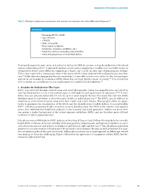
Table 5: Findings on glaucoma examination that warrant investigation into other differential diagnoses: 176
Test Results
• Presenting BCVA <20/40
• Age <50 years
• + RAPD
• Optic nerve pallor
• Neurological symptoms
(headaches, weakness, numbness, etc.)
• Visual field defects respecting vertical midline
• Abnormal progression of visual field defects
To properly assess the optic nerve, it is critical to define the NRR by contour, noting the deflection of fine blood
vessels, rather than pallor. A mismatch between central pallor (suggesting a ‘smaller cup’) and NRR margin as
40
delineated by blood vessel deflection (suggesting a ‘larger cup’) can be an early sign of glaucomatous damage.
This is best noted with a stereoscopic view of the nerve, which is best obtained with a dilated fundus examina-
tion. While objective imaging has become invaluable, it is not able to detect rim pallor (or disc hemorrhages)
44
and can be confounded by anomalous ONHs (those that are tilted, highly myopic, or pitted). It is critical that
177
OCT is viewed as a complement to, not a replacement for careful clinical evaluation. 178
3. Examine the Retinal nerve fiber layer
RNFL loss detected through clinical exam and serial photography (using low magnification and aided by
red-free illumination) is one of the earliest signs of, although not pathognomonic for glaucoma. 179,180 In fact,
RNFL loss can precede detectable VF loss by up to 6 years despite the fact that more than half the RNFL
thickness must be lost before a defect becomes visible on ophthalmoscopy. The RNFL may be difficult to
181
visualize on clinical examination, even with clear media and a dark fundus. Photography offers an oppor-
tunity to maximize the visualization of the RNFL and the identification of subtle defects. A normal healthy
RNFL will show prominent bright striations as nerve bundles enter the ONH at the inferior and superior
poles, with relatively less brightness adjacent to the temporal and nasal quadrants. Defects are more obvi-
ous against a darker background of the retinal pigment epithelium (RPE), and are therefore more difficult
to detect in lightly pigmented eyes.
Like glaucomatous NRR defects, RNFL defects can be either diffuse or focal. Diffuse thinning dulls the normally
bright RNFL striations, enhances visibility of the parapapillary retinal vessels, and typically manifests as asym-
metry between superior and inferior hemispheres and between right and left eyes. One should pay particular
182
attention to any asymmetries in brightness or blood vessel clarity between the eyes, as well as between the supe-
rior and inferior poles of the optic nerve head. Diffuse glaucomatous loss is superimposed on diffuse age-related
loss, making its detection challenging. Figure 5 illustrates the appearance of asymmetric diffuse RNFL loss be-
tween the right and left eye.
CANADIAN JOURNAL of OPTOMETRY | REVUE CANADIENNE D’OPTOMÉTRIE VOL. 79 SUPPLEMENT 1, 2017 23