
Page 24 - CJO_F17_GLAUCOMA_SUPPLEMENT
P. 24
C CLINICAL RESEARCH
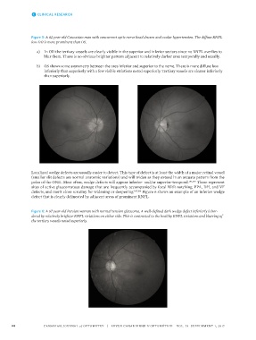
Figure 5: A 62 year-old Caucasian man with concurrent optic nerve head drusen and ocular hypertension. The diffuse RNFL
loss OD is more prominent than OS.
a) In OD the tertiary vessels are clearly visible in the superior and inferior sectors since no RNFL overlies to
blur them. There is no obvious brighter pattern adjacent to relatively darker area temporally and nasally.
b) OS shows some asymmetry between the area inferior and superior to the nerve. There is more diffuse loss
inferiorly than superiorly with a few visible striations noted superiorly. Tertiary vessels are clearer inferiorly
than superiorly.
Localized wedge defects are usually easier to detect. This type of defect is at least the width of a major retinal vessel
(smaller slit defects are normal anatomic variations) and will widen as they extend in an arcuate pattern from the
poles of the ONH. Most often, wedge defects will appear inferior- and/or superior-temporal. 183,184 These represent
sites of active glaucomatous damage that are frequently accompanied by focal NRR notching, PPA, DH, and VF
defects, and merit close scrutiny for widening or deepening. 185,186 Figure 6 shows an example of an inferior wedge
defect that is clearly delineated by adjacent areas of prominent RNFL.
Figure 6: A 67 year-old Persian woman with normal tension glaucoma. A well-defined dark wedge defect inferiorly is bor-
dered by relatively brighter RNFL striations on either side. This is contrasted to the healthy RNFL striations and blurring of
the tertiary vessels noted superiorly.
24 CANADIAN JOURNAL of OPTOMETRY | REVUE CANADIENNE D’OPTOMÉTRIE VOL. 79 SUPPLEMENT 1, 2017