
Page 267 - Anatomy and Physiology of Farm Animals, 8th Edition
P. 267
252 / Anatomy and Physiology of Farm Animals
the retina. Most of the axons of the optic Many domestic species are seasonal
nerve synapse in the thalamus, and from
VetBooks.ir there visual information travels to the breeders, meaning that their repro-
ductive cycles are determined by the
primary visual cortex in the occipital
lobe of the brain (the most caudal part of the season. The most powerful determi-
nant of the onset and cessation of
cerebral cortex) for conscious perception. breeding cycles in these species is the
A smaller subset of ganglion cell axons length of the day. The retinal projec-
project to other destinations in the brain. tions to the suprachiasmatic nucleus
Some reach the rostral colliculi of the are the brain’s record of day length,
mesencephalon, where visual stimuli and they therefore determine the
induce reflex movements of the eyes and reproductive cycles via their influence
head. Others project to the pretectal on the autonomic functions of the hypo-
nuclei, also in the region of the mesen thalamus. It is common agricultural
cephalon; these nuclei communicate with practice to alter breeding behavior by
the oculomotor nuclei to coordinate the exposing animals to artificial light.
reflex constriction of the pupils in response For instance, in the horse industry, in
to light. Finally, a very small number of which an early foaling date is desira-
ganglion cell axons project to a specific ble, mares are commonly exposed to
group of cells of the hypothalamus, the artificially increased day length in the
suprachiasmatic nucleus. The suprachi winter so as to cause these spring
asmatic nucleus is the biologic clock, the breeders to begin fertile estrous cycles
part of the brain that sets circadian earlier than they would if exposed only
rhythms. Circadian rhythms are physio to natural light.
logic processes that vary regularly on a There is a widely repeated myth among
daily basis; prominent circadian rhythms horse trainers that states that visual
include sleep–wake cycles, melatonin information from one side of the body is
secretion, and body temperature fluctua processed strictly on the opposite side of
tions. The suprachiasmatic nucleus has the brain and vice versa. Horse trainers
an intrinsic rhythmicity that closely have often cited this “fact” as a rationale
approximates 24 hours, but the projec for schooling horses from both sides of
tions from the retina keeps the nucleus’s the body. While it is true that herbivores
cycle entrained to the actual photoperiod like horses process a majority of visual
of the day (Fig. 12‐16). information from each half of their visual
field in the contralateral visual cortex,
there are three reasons why this idea is
not neurobiologically sound. One is that
not all of the visual information from
a each eye crosses to the contralateral cor
tex; some is processed on the ipsilateral
b
c (same) side. Secondly, a small portion of
d the equine visual field is binocular, that
is, is seen simultaneously by both eyes.
And finally, the caudal part of the corpus
callosum (the large bundle of axons
connecting right and left cerebral hemi
spheres) connects the visual cortices of
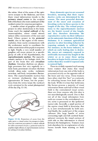
Figure 12-16. Projections of axons from the
optic nerve. a, visual cortex (via synapse in thala each side so that information is shared
mus) for conscious perception; b, rostral colliculi between the hemispheres. Trainers must
for reflex movement of eyes; c, pretectal nuclei for look elsewhere for an explanation of why
pupillary light reflexes; d, suprachiasmatic nucleus horses benefit from training on both
for setting of circadian rhythms. sides.