
Page 264 - Anatomy and Physiology of Farm Animals, 8th Edition
P. 264
Sense Organs / 249
(A) (B)
VetBooks.ir
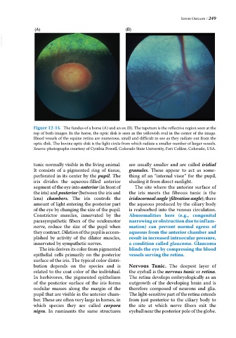
Figure 12-14. The fundus of a horse (A) and an ox (B). The tapetum is the reflective region seen at the
top of both images. In the horse, the optic disk is seen as the yellowish oval in the center of the image.
Blood vessels of the equine retina are numerous, small and difficult to see as they radiate out from the
optic disk. The bovine optic disk is the light circle from which radiate a smaller number of larger vessels.
Source: photographs courtesy of Cynthia Powell, Colorado State University, Fort Collins, Colorado, USA.
tunic normally visible in the living animal. are usually smaller and are called iridial
It consists of a pigmented ring of tissue, granules. These appear to act as some
perforated in its center by the pupil. The thing of an “internal visor” for the pupil,
iris divides the aqueous‐filled anterior shading it from direct sunlight.
segment of the eye into anterior (in front of The site where the anterior surface of
the iris) and posterior (between the iris and the iris meets the fibrous tunic is the
lens) chambers. The iris controls the iridocorneal angle (filtration angle); there
amount of light entering the posterior part the aqueous produced by the ciliary body
of the eye by changing the size of the pupil. is reabsorbed into the venous circulation.
Constrictor muscles, innervated by the Abnormalities here (e.g., congenital
parasympathetic fibers of the oculomotor narrowing or obstruction due to inflam-
nerve, reduce the size of the pupil when mation) can prevent normal egress of
they contract. Dilation of the pupil is accom aqueous from the anterior chamber and
plished by activity of the dilator muscles, result in increased intraocular pressure,
innervated by sympathetic nerves. a condition called glaucoma. Glaucoma
The iris derives its color from pigmented blinds the eye by compressing the blood
epithelial cells primarily on the posterior vessels serving the retina.
surface of the iris. The typical color distri
bution depends on the species and is Nervous Tunic. The deepest layer of
related to the coat color of the individual. the eyeball is the nervous tunic or retina.
In herbivores, the pigmented epithelium The retina develops embryologically as an
of the posterior surface of the iris forms outgrowth of the developing brain and is
nodular masses along the margin of the therefore composed of neurons and glia.
pupil that are visible in the anterior cham The light‐sensitive part of the retina extends
ber. These are often very large in horses, in from just posterior to the ciliary body to
which species they are called corpora the site at which nerve fibers exit the
nigra. In ruminants the same structures eyeball near the posterior pole of the globe.