
Page 107 - Essential Haematology
P. 107
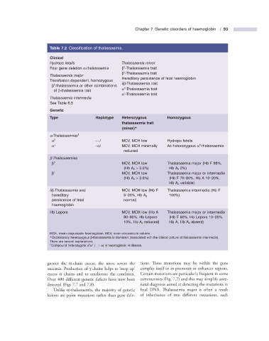
Chapter 7  Genetic disorders of haemoglobin   /  93


                            Table 7.2   Classifi cation of thalassaemia.

                                Clinical
                                Hydrops fetalis               Thalassaemia minor
                                                         0


                              Four gene deletion  α - thalassaemia         β  - Thalassaemia trait
                                                           +

                                                                β  - Thalassaemia trait

                                Thalassaemia major
                                                              Hereditary persistence of fetal haemoglobin
                              Transfusion dependent, homozygous

                                                                δ β - Thalassaemia trait

                         0

                                β  - thalassaemia or other combinations

                                                         0

                                                                α  - Thalassaemia trait

                         of  β - thalassaemia trait

                                                           +
                                                                α  - Thalassaemia trait


                                Thalassaemia intermedia
                              See Table  6.5
                                Genetic
                            Type              Haplotype     Heterozygous         Homozygous
                                                        thalassaemia trait

                                                        (minor) *
                                      †


                                α - Thalassaemias
                         0

                                α                 – – /     MCV, MCH low       Hydrops fetalis


                                                                                           0
                           +


                                α               – α /         MCV, MCH minimally       As heterozygous  α  - thalassaemia


                                                         reduced
                                β - Thalassaemias

                         0
                                β                         MCV, MCH low           Thalassaemia major (Hb F 98%,


                                                         (Hb A  2   >  3.5%)    Hb A  2  2%)
                           +
                                β                         MCV, MCH low           Thalassaemia major or intermedia


                                                         (Hb A  2    >  3.5%)    (Hb F 70 – 80%, Hb A 10 – 20%,
                                                                             Hb A  2   variable)


                              δ β - Thalassaemia and            MCV, MCH low (Hb F       Thalassaemia intermedia (Hb F
                        hereditary                       5 – 20%, Hb A  2      100%)
                        persistence of fetal             normal)
                        haemoglobin
                              Hb Lepore                     MCV, MCH low (Hb A         Thalassaemia major or intermedia
                                                         80 – 90%, Hb Lepore   (Hb F 80%, Hb Lepore 10 – 20%,
                                                         10%, Hb A  2   reduced)    Hb A, Hb A  2   absent)
                              MCH, mean corpuscular haemoglobin; MCV, mean corpuscular volume.


                              *   Occasionally heterozygous  β - thalassaemia is dominant (associated with the clinical picture of thalassaemia intermedia).
                        There are several explanations.
                          †              0    +


                                Compound heterozygote  α  α   ( – – / – α ) is haemoglobin H disease.


                      greater the  α - chain excess, the more severe the   tions. These mutations may be within the gene

                                                       ‘
                      anaemia. Production of  γ  chains helps to   mop up ’    complex itself or in promoter or enhancer regions.
                      excess  α  chains and to ameliorate the condition.   Certain mutations are particularly frequent in some
                      Over 400 different genetic defects have now been   communities (Fig.  7.7 ) and this may simplify ante-

                      detected (Figs  7.7  and  7.8 ).          natal diagnosis aimed at detecting the mutations in


                          Unlike  α - thalassaemia, the majority of genetic   fetal DNA. Thalassaemia major is often a result

                      lesions are point mutations rather than gene dele-  of inheritance of two different mutations, each