
Page 108 - Essential Haematology
P. 108
94 / Chapter 7 Genetic disorders of haemoglobin
(a) (b)
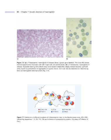
Figure 7.6 (a) α - Thalassaemia: haemoglobin H disease (three α - globin gene deletion). The blood fi lm shows
marked hypochromic microcytic cells with target cells and poikilocytosis. (b) α - Thalassaemia: haemoglobin H
disease. Supravital staining with brilliant cresyl blue reveals multiple fi ne, deeply stained deposits ( ‘ golf ball ’
cells) caused by precipitation of aggregates of β - globin chains. Hb H can also be detected as a fast - moving
band on haemoglobin electrophoresis (Fig. 7.12 ).
+
+
0
β IVS1-110 β 39 β IVS2-745
+
0
β IVS1-1 β IVS1-6 Others
Figure 7.7 Distribution of different mutations of β - thalassaemia major in the Mediterranean area. IVSI, IVS2
intervening sequences; 1, 6, 39, 110, 745 are mutations of corresponding codons. (Courtesy of Professor A.
Cao.)