
Page 109 - Essential Haematology
P. 109
Chapter 7 Genetic disorders of haemoglobin / 95
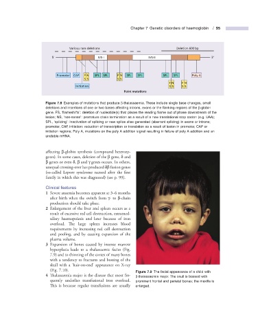
Various rare deletions Deletion 600 bp
5' IVS-I IVS-II 3'
Promoter CAP F N SPL SPL F N SPL SPL SPL SPL Poly A
S S S S
F N F N
Initiation S S S S
Point mutations
Figure 7.8 Examples of mutations that produce β - thalassaemia. These include single base changes, small
deletions and insertions of one or two bases affecting introns, exons or the fl anking regions of the β - globin
gene. FS, ‘ frameshifts ’ : deletion of nucleotide(s) that places the reading frame out of phase downstream of the
lesion; NS, ‘ non - sense ’ : premature chain termination as a result of a new translational stop codon (e.g. UAA);
SPL, ‘ splicing ’ : inactivation of splicing or new splice sites generated (aberrant splicing) in exons or introns;
promoter, CAP, initiation: reduction of transcription or translation as a result of lesion in promoter, CAP or
initiation regions; Poly A, mutations on the poly A addition signal resulting in failure of poly A addition and an
unstable mRNA.
aff ecting β - globin synthesis (compound heterozy-
gotes). In some cases, deletion of the β gene, δ and
β genes or even δ , β and γ genes occurs. In others,
unequal crossing - over has produced δ β fusion genes
(so - called Lepore syndrome named after the fi rst
family in which this was diagnosed) (see p. 99) .
Clinical f eatures
1 Severe anaemia becomes apparent at 3 – 6 months
after birth when the switch from γ - to β - chain
production should take place.
2 Enlargement of the liver and spleen occurs as a
result of excessive red cell destruction, extramed-
ullary haemopoiesis and later because of iron
overload. The large spleen increases blood
requirements by increasing red cell destruction
and pooling, and by causing expansion of the
plasma volume.
3 Expansion of bones caused by intense marrow
hyperplasia leads to a thalassaemic facies (Fig.
7.9 ) and to thinning of the cortex of many bones
with a tendency to fractures and bossing of the
skull with a ‘ hair - on - end ’ appearance on X - ray
(Fig. 7.10 ). Figure 7.9 The facial appearance of a child with
4 Thalassaemia major is the disease that most fre- β - thalassaemia major. The skull is bossed with
quently underlies transfusional iron overload. prominent frontal and parietal bones; the maxilla is
This is because regular transfusions are usually enlarged.