
Page 114 - Essential Haematology
P. 114
100 / Chapter 7 Genetic disorders of haemoglobin
Vaso - o cclusive c rises
Amino acid pro glu glu
Normal β-chain These are the most frequent and are precipitated by
Base composition CCT G A G GAG
such factors as infection, acidosis, dehydration or
Base composition CCT G T G GAG deoxygenation (e.g. altitude, operations, obstetric
Sickle β-chain delivery, stasis of the circulation, exposure to cold,
Amino acid pro val glu
violent exercise). Infarcts causing severe pain occur
in the bones (hips, shoulders and vertebrae are com-
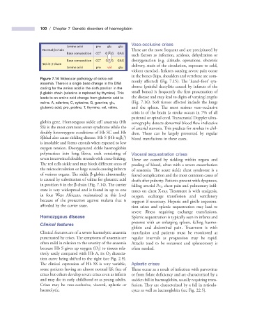
Figure 7.14 Molecular pathology of sickle cell
monly affected) (Fig. 7.15 ). The ‘ hand – foot ’ syn-
anaemia. There is a single base change in the DNA
drome (painful dactylitis caused by infarcts of the
coding for the amino acid in the sixth position in the
small bones) is frequently the first presentation of
β - globin chain (adenine is replaced by thymine). This
leads to an amino acid change from glutamic acid to the disease and may lead to digits of varying lengths
valine. A, adenine; C, cytosine; G, guanine; glu, (Fig. 7.16 ). Soft tissues affected include the lungs
glutamic acid; pro, proline; T, thymine; val, valine. and the spleen. The most serious vaso - occlusive
crisis is of the brain (a stroke occurs in 7% of all
patients) or spinal cord. Transcranial Doppler ultra-
globin gene. Homozygous sickle cell anaemia (Hb sonography detects abnormal blood fl ow indicative
SS) is the most common severe syndrome while the of arterial stenosis. This predicts for strokes in chil-
doubly heterozygote conditions of Hb SC and Hb dren. These can be largely prevented by regular
S
S β thal also cause sickling disease. Hb S (Hb α 2 β 2 ) blood transfusions in these cases.
is insoluble and forms crystals when exposed to low
oxygen tension. Deoxygenated sickle haemoglobin
polymerizes into long fibres, each consisting of Visceral s equestration c rises
seven intertwined double strands with cross - linking. These are caused by sickling within organs and
The red cells sickle and may block different areas of pooling of blood, often with a severe exacerbation
the microcirculation or large vessels causing infarcts of anaemia. The acute sickle chest syndrome is a
of various organs. The sickle β - globin abnormality feared complication and the most common cause of
is caused by substitution of valine for glutamic acid death after puberty. Patients present with dyspnoea,
in position 6 in the β chain (Fig. 7.14 ). Th e carrier falling arterial P O 2 , chest pain and pulmonary infi l-
state is very widespread and is found in up to one trates on chest X - ray. Treatment is with analgesia,
in four West Africans, maintained at this level oxygen, exchange transfusion and ventilatory
because of the protection against malaria that is support if necessary. Hepatic and girdle sequestra-
afforded by the carrier state. tion crises and splenic sequestration may lead to
severe illness requiring exchange transfusions.
Homozygous d isease Splenic sequestration is typically seen in infants and
presents with an enlarging spleen, falling haemo-
Clinical f eatures
globin and abdominal pain. Treatment is with
Clinical features are of a severe haemolytic anaemia transfusion and patients must be monitored at
punctuated by crises. The symptoms of anaemia are regular intervals as progression may be rapid.
often mild in relation to the severity of the anaemia Attacks tend to be recurrent and splenectomy is
because Hb S gives up oxygen (O 2 ) to tissues rela- often needed.
tively easily compared with Hb A, its O 2 dissocia-
tion curve being shifted to the right (see Fig. 2.9 ).
The clinical expression of Hb SS is very variable, Aplastic c rises
some patients having an almost normal life free of Th ese occur as a result of infection with parvovirus
crises but others develop severe crises even as infants or from folate defi ciency and are characterized by a
and may die in early childhood or as young adults. sudden fall in haemoglobin, usually requiring trans-
Crises may be vaso - occlusive, visceral, aplastic or fusion. Th ey are characterized by a fall in reticulo-
haemolytic. cytes as well as haemoglobin (see Fig. 22.5 ).