
Page 115 - Essential Haematology
P. 115
Chapter 7 Genetic disorders of haemoglobin / 101
(a)
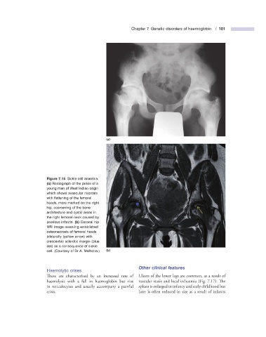
Figure 7.15 Sickle cell anaemia.
(a) Radiograph of the pelvis of a
young man of West Indian origin
which shows avascular necrosis
with fl attening of the femoral
heads, more marked on the right
hip, coarsening of the bone
architecture and cystic areas in
the right femoral neck caused by
previous infarcts. (b) Coronal hip
MRI image revealing established
osteonecrosis of femoral heads
bilaterally (yellow arrow) with
crescentric sclerotic margin (blue
dot) as a consequence of sickle
cell. (Courtesy of Dr A. Malhotra.) (b)
Other c linical f eatures
Haemolytic c rises
These are characterized by an increased rate of Ulcers of the lower legs are common, as a result of
haemolysis with a fall in haemoglobin but rise vascular stasis and local ischaemia (Fig. 7.17 ). Th e
in reticulocytes and usually accompany a painful spleen is enlarged in infancy and early childhood but
crisis. later is often reduced in size as a result of infarcts