
Page 120 - Essential Haematology
P. 120
106 / Chapter 7 Genetic disorders of haemoglobin
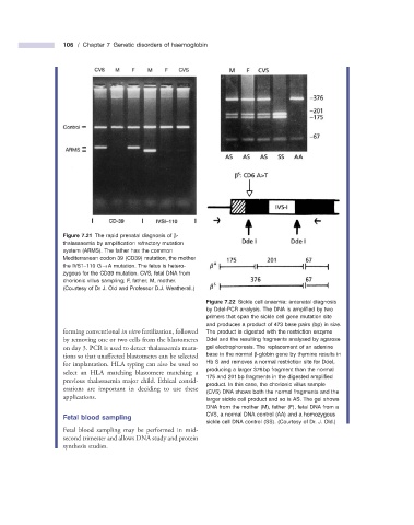
Figure 7.21 The rapid prenatal diagnosis of β -
thalassaemia by amplifi cation refractory mutation
system (ARMS). The father has the common
Mediterranean codon 39 (CD39) mutation, the mother
the IVS1 – 110 G → A mutation. The fetus is hetero-
zygous for the CD39 mutation. CVS, fetal DNA from
chorionic villus sampling; F, father; M, mother.
(Courtesy of Dr J. Old and Professor D.J. Weatherall.)
Figure 7.22 Sickle cell anaemia: antenatal diagnosis
by DdeI - PCR analysis. The DNA is amplifi ed by two
primers that span the sickle cell gene mutation site
and produces a product of 473 base pairs (bp) in size.
forming conventional in vitro fertilization, followed The product is digested with the restriction enzyme
by removing one or two cells from the blastomeres DdeI and the resulting fragments analysed by agarose
on day 3. PCR is used to detect thalassaemia muta- gel electrophoresis. The replacement of an adenine
tions so that unaffected blastomeres can be selected base in the normal β - globin gene by thymine results in
for implantation. HLA typing can also be used to Hb S and removes a normal restriction site for DdeI,
producing a larger 376 bp fragment than the normal
select an HLA matching blastomere matching a
175 and 201 bp fragments in the digested amplifi ed
previous thalassaemia major child. Ethical consid-
product. In this case, the chorionic villus sample
erations are important in deciding to use these
(CVS) DNA shows both the normal fragments and the
applications. larger sickle cell product and so is AS. The gel shows
DNA from the mother (M), father (F), fetal DNA from a
Fetal b lood s ampling CVS, a normal DNA control (AA) and a homozygous
sickle cell DNA control (SS). (Courtesy of Dr. J. Old.)
Fetal blood sampling may be performed in mid -
second trimester and allows DNA study and protein
synthesis studies.