
Page 131 - Essential Haematology
P. 131
Chapter 8 White cells: Granulocytes and monocytes / 117
mild thrombocytopenia with giant platelets.
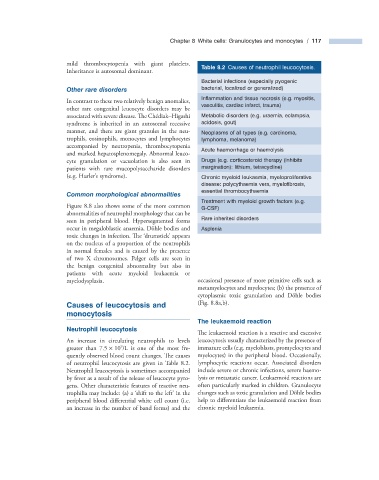
Table 8.2 Causes of neutrophil leucocytosis.
Inheritance is autosomal dominant.
Bacterial infections (especially pyogenic
Other r are d isorders bacterial, localized or generalized)
Infl ammation and tissue necrosis (e.g. myositis,
In contrast to these two relatively benign anomalies,
vasculitis, cardiac infarct, trauma)
other rare congenital leucocyte disorders may be
associated with severe disease. The Ch é diak – Higashi Metabolic disorders (e.g. uraemia, eclampsia,
syndrome is inherited in an autosomal recessive acidosis, gout)
manner, and there are giant granules in the neu- Neoplasms of all types (e.g. carcinoma,
trophils, eosinophils, monocytes and lymphocytes lymphoma, melanoma)
accompanied by neutropenia, thrombocytopenia
Acute haemorrhage or haemolysis
and marked hepatosplenomegaly. Abnormal leuco-
cyte granulation or vacuolation is also seen in Drugs (e.g. corticosteroid therapy (inhibits
patients with rare mucopolysaccharide disorders margination): lithium, tetracycline)
(e.g. Hurler ’ s syndrome). Chronic myeloid leukaemia, myeloproliferative
disease: polycythaemia vera, myelofi brosis,
essential thrombocythaemia
Common m orphological a bnormalities
Treatment with myeloid growth factors (e.g.
Figure 8.8 also shows some of the more common G - CSF)
abnormalities of neutrophil morphology that can be
seen in peripheral blood. Hypersegmented forms Rare inherited disorders
occur in megaloblastic anaemia, D ö hle bodies and Asplenia
toxic changes in infection. The ‘ drumstick ’ appears
on the nucleus of a proportion of the neutrophils
in normal females and is caused by the presence
of two X chromosomes. Pelger cells are seen in
the benign congenital abnormality but also in
patients with acute myeloid leukaemia or
myelodysplasia. occasional presence of more primitive cells such as
metamyelocytes and myelocytes; (b) the presence of
cytoplasmic toxic granulation and D ö hle bodies
Causes of l eucocytosis and (Fig. 8.8 a,b).
m onocytosis
The l eukaemoid r eaction
Neutrophil l eucocytosis
The leukaemoid reaction is a reactive and excessive
An increase in circulating neutrophils to levels leucocytosis usually characterized by the presence of
9
greater than 7.5 × 10 /L is one of the most fre- immature cells (e.g. myeloblasts, promyelocytes and
quently observed blood count changes. Th e causes myelocytes) in the peripheral blood. Occasionally,
of neutrophil leucocytosis are given in Table 8.2 . lymphocytic reactions occur. Associated disorders
Neutrophil leucocytosis is sometimes accompanied include severe or chronic infections, severe haemo-
by fever as a result of the release of leucocyte pyro- lysis or metastatic cancer. Leukaemoid reactions are
gens. Other characteristic features of reactive neu- often particularly marked in children. Granulocyte
trophilia may include: (a) a shift to the left ’ in the changes such as toxic granulation and D ö hle bodies
‘
peripheral blood differential white cell count (i.e. help to diff erentiate the leukaemoid reaction from
an increase in the number of band forms) and the chronic myeloid leukaemia.