
Page 130 - Essential Haematology
P. 130
116 / Chapter 8 White cells: Granulocytes and monocytes
Phagocytosis leukaemia and myelodysplastic syndromes may also
be associated with defective killing of ingested
These defects usually arise because of a lack of
microorganisms.
opsonization which may be caused by congenital or
acquired causes of hypogammaglobulinaemia or
lack of complement components. Benign d isorders
A number of the hereditary conditions may give rise
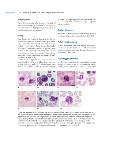
Killing to changes in granulocyte morphology (Fig. 8.8 ).
This abnormality is clearly illustrated by the rare
X - linked or autosomal recessive chronic granuloma- Pelger – Hu ë t a nomaly
tous disease that results from abnormal leucocyte
oxidative metabolism. There is an abnormality In this uncommon condition bilobed neutrophils
aff ecting different elements of the respiratory burst are found in the peripheral blood. Occasional
oxidase or its activating mechanism. Th e patients unsegmented neutrophils are also seen. Inheritance
have recurring infections, usually bacterial but is autosomal dominant.
sometimes fungal, which present in infancy or early
childhood in most cases.
May – Hegglin a nomaly
Other rare congenital abnormalities may also
result in defects of bacterial killing (e.g. myeloper- In this rare condition the neutrophils contain
oxidase deficiency and the Ch é diak – Higashi syn- basophilic inclusions of RNA (resembling D ö hle
drome; see below). Acute or chronic myeloid bodies) in the cytoplasm. There is an associated
(a) (b) (c) (d)
(e) (f) (g)
Figure 8.8 Abnormal white blood cells. (a) Neutrophil leucocytosis: toxic changes shown by the presence of
red – purple granules in the band form neutrophils. (b) Neutrophil leucocytosis: a D ö hle body can be seen in the
cytoplasm of the neutrophil. (c) Megaloblastic anaemia: hypersegmented oversized neutrophil in peripheral
blood. (d) May – Hegglin anomaly: the neutrophils contain basophilic inclusions 2 – 5 μ m in diameter; there is an
associated mild thrombocytopenia with giant platelets. (e) Pelger – Hu ë t anomaly: coarse clumping of the
chromatin in pince nez confi guration. (f) Ch é diak – Higashi syndrome: bizarre giant granules in the cytoplasm of
a monocyte. (g) Alder ’ s anomaly: coarse violet granules in the cytoplasm of a neutrophil.