
Page 135 - Essential Haematology
P. 135
Chapter 8 White cells: Granulocytes and monocytes / 121
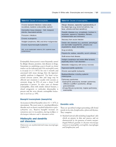
Table 8.5 Causes of monocytosis. Table 8.6 Causes of eosinophilia.
Chronic bacterial infections: tuberculosis, Allergic diseases, especially hypersensitivity of
brucellosis, bacterial endocarditis, typhoid the atopic type (e.g. bronchial asthma, hay
fever, urticaria and food sensitivity)
Connective tissue diseases – SLE, temporal
arteritis, rheumatoid arthritis Parasitic diseases (e.g. amoebiasis, hookworm,
ascariasis, tapeworm infestation, fi lariasis,
Protozoan infections
schistosomiasis and trichinosis)
Chronic neutropenia
Recovery from acute infection
Hodgkin lymphoma, AML and other malignancies
Certain skin diseases (e.g. psoriasis, pemphigus
Chronic myelomonocytic leukaemia and dermatitis herpetiformis, urticaria and
angioedema, atopic dermatitis)
AML, acute myeloblastic leukemia; SLE, systemic lupus Drug sensitivity
erythematosus.
Polyarteritis nodosa, vasculitis, serum sickness
Graft - versus - host disease
Hodgkin lymphoma and some other tumours,
Eosinophilic leucocytosis is most frequently caused especially clonal T - cell disorders
by allergic diseases, parasites, skin diseases or drugs.
Metastatic malignancy with tumour necrosis
Sometimes no underlying cause is found, no clonal
marker can be indicated and if the eosinophil count Hypereosinophilic syndrome
9
is elevated ( > 1.5 × 10 /L) for over 6 months and Chronic eosinophilic leukaemia
associated with tissue damage then the hypereosi-
Myeloproliferative including systemic
nophilic syndrome is diagnosed. The heart valves,
mastocytosis
central nervous system, skin and lungs may be
affected and treatment is usually with steroids or Pulmonary syndromes
cytotoxic drugs. In 25% of cases a clonal T - cell Eosinophilic pneumonia, transient pulmonary
population is present. In other cases of chronic infi ltrates (Loeffl er ’ s syndrome), allergic
granulomatosis
eosinophilia, often with similar clinical features, a
(Churg – Strauss syndrome), tropical pulmonary
clonal cytogenetic or molecular abnormality is
eosinophilia
present and the term chronic eosinophilic leukae-
mia is used (see p. 199) .
Basophil l eucocytosis ( b asophilia)
9
An increase in blood basophils above 0.1 × 10 /L is
uncommon. The usual cause is a myeloproliferative Dendritic c ells
disorder such as chronic myeloid leukaemia or poly- These are specialized antigen - presenting cells found
cythaemia vera. Reactive basophil increases are mainly in the skin, lymph nodes, spleen and thymus.
sometimes seen in myxoedema, during smallpox or Th ey comprise:
chickenpox infection and in ulcerative colitis.
1 Myeloid - derived cells including Langerhans ’ cells
Histiocytic and d endritic which are present in skin and mucosa and are
characterized by the presence of tennis racquet -
c ell d isorders
shaped Birbeck granules in electron - microscopy
Histiocytes are myeloid - derived tissue macrophages sections in neutrophils, eosinophils, macrophages
(Table 8.7 ). and lymphocytes; and