
Page 152 - Essential Haematology
P. 152
138 / Chapter 9 White cells: Lymphocytes
(a) (b)
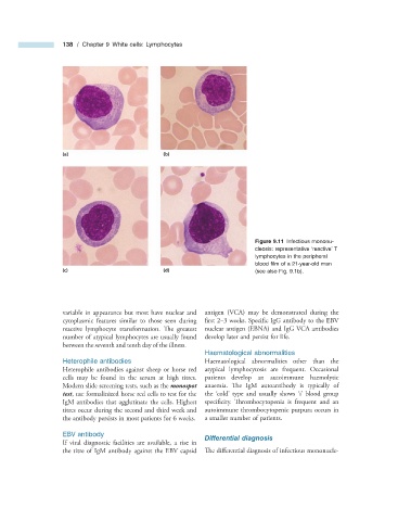
Figure 9.11 Infectious mononu-
cleosis: representative ‘ reactive ’ T
lymphocytes in the peripheral
blood fi lm of a 21 - year - old man
(c) (d) (see also Fig. 9.1 b).
variable in appearance but most have nuclear and antigen (VCA) may be demonstrated during the
cytoplasmic features similar to those seen during first 2 – 3 weeks. Specific IgG antibody to the EBV
reactive lymphocyte transformation. Th e greatest nuclear antigen (EBNA) and IgG VCA antibodies
number of atypical lymphocytes are usually found develop later and persist for life.
between the seventh and tenth day of the illness.
Haematological a bnormalities
Heterophile a ntibodies Haematological abnormalities other than the
Heterophile antibodies against sheep or horse red atypical lymphocytosis are frequent. Occasional
cells may be found in the serum at high titres. patients develop an autoimmune haemolytic
Modern slide screening tests, such as the monospot anaemia. The IgM autoantibody is typically of
test , use formalinized horse red cells to test for the the ‘ cold ’ type and usually shows ‘ i ’ blood group
IgM antibodies that agglutinate the cells. Highest specifi city. Thrombocytopenia is frequent and an
titres occur during the second and third week and autoimmune thrombocytopenic purpura occurs in
the antibody persists in most patients for 6 weeks. a smaller number of patients.
EBV a ntibody
Differential d iagnosis
If viral diagnostic facilities are available, a rise in
the titre of IgM antibody against the EBV capsid Th e differential diagnosis of infectious mononucle-