Page 151 - Essential Haematology
P. 151

Chapter 9  White cells: Lymphocytes  /  137


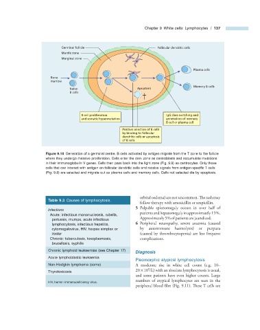
                              Germinal follicle                            Follicular dendritic cells
                              Mantle zone
                              Marginal zone

                                                                                           Plasma cells
                         Bone
                         marrow
                                                                                           Memory B cells
                                  Naive                          Apoptosis
                                  B cells


                                       B cell proliferation                    IgG class switching and
                                       and somatic hypermutation               generation of memory
                                                                               B cell or plasma cell
                                                          Positive selection of B cells
                                                          by binding to follicular
                                                          dendritic cells or apoptosis
                                                          of B cells

                                Figure 9.10   Generation of a germinal centre. B cells activated by antigen migrate from the T zone to the follicle


                      where they undergo massive proliferation. Cells enter the dark zone as centroblasts and accumulate mutations
                      in their immunoglobulin V genes. Cells then pass back into the light zone (Fig.  9.9 ) as centrocytes. Only those
                      cells that can interact with antigen on follicular dendritic cells and receive signals from antigen - specifi c T cells
                      (Fig.  9.8 ) are selected and migrate out as plasma cells and memory cells. Cells not selected die by apoptosis.


                                                                  orbital oedema) are not uncommon. The rash may


                            Table 9.3   Causes of lymphocytosis.
                                                                  follow therapy with amoxicillin or ampicillin.
                                                                   5      Palpable splenomegaly occurs in over half of
                              Infections
                                                                  patients and hepatomegaly in approximately 15%.
                                Acute: infectious mononucleosis, rubella,
                         pertussis, mumps, acute infectious       Approximately 5% of patients are jaundiced.
                         lymphocytosis, infectious hepatitis,      6      Peripheral  neuropathy,  severe  anaemia  (caused
                         cytomegalovirus, HIV, herpes simplex or   by autoimmune haemolysis) or purpura
                         zoster                                   (caused by thrombocytopenia) are less frequent
                                Chronic: tuberculosis, toxoplasmosis,   complications.
                         brucellosis, syphilis

                            Chronic lymphoid leukaemias (see Chapter  17 )         Diagnosis

                            Acute lymphoblastic leukaemia
                                                                    Pleomorphic  a typical  l ymphocytosis
                            Non - Hodgkin lymphoma (some)         A moderate rise in white cell count (e.g. 10 –
                                                                      9
                                                                 20    ×    10  /L) with an absolute lymphocytosis is usual,
                            Thyrotoxicosis
                                                                and some patients have even higher counts. Large
                                                                numbers of atypical lymphocytes are seen in the
                              HIV, human immunodefi ciency virus.

                                                                peripheral blood film (Fig.  9.11 ). These T cells are
   146   147   148   149   150   151   152   153   154   155   156