
Page 194 - Essential Haematology
P. 194
180 / Chapter 13 Acute myeloid leukaemia
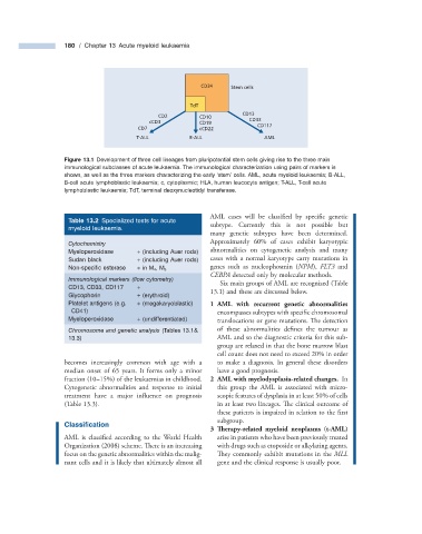
CD34 Stem cells
TdT
CD13
CD2 CD10
cCD3 CD19 CD33
CD7 cCD22 CD117
T-ALL B-ALL AML
Figure 13.1 Development of three cell lineages from pluripotential stem cells giving rise to the three main
immunological subclasses of acute leukaemia. The immunological characterization using pairs of markers is
shown, as well as the three markers characterizing the early ‘ stem ’ cells. AML, acute myeloid leukaemia; B - ALL,
B - cell acute lymphoblastic leukaemia; c, cytoplasmic; HLA, human leucocyte antigen; T - ALL, T - cell acute
lymphoblastic leukaemia; TdT, terminal deoxynucleotidyl transferase.
AML cases will be classified by specifi c genetic
Table 13.2 Specialized tests for acute
myeloid leukaemia. subtype. Currently this is not possible but
many genetic subtypes have been determined.
Cytochemistry Approximately 60% of cases exhibit karyotypic
Myeloperoxidase + (including Auer rods) abnormalities on cytogenetic analysis and many
Sudan black + (including Auer rods) cases with a normal karyotype carry mutations in
Non - specifi c esterase + in M 4 , M 5 genes such as nucleophosmin ( NPM ), FLT3 and
CEBPA detected only by molecular methods.
Immunological markers (fl ow cytometry)
Six main groups of AML are recognized (Table
CD13, CD33, CD117 +
13.1 ) and these are discussed below.
Glycophorin + (erythroid)
Platelet antigens (e.g. + (megakaryoblastic) 1 AML with recurrent genetic abnormalities
CD41) encompasses subtypes with specifi c chromosomal
Myeloperoxidase + (undifferentiated) translocations or gene mutations. Th e detection
Chromosome and genetic analysis (Tables 13.1 & of these abnormalities defines the tumour as
13.3) AML and so the diagnostic criteria for this sub-
group are relaxed in that the bone marrow blast
cell count does not need to exceed 20% in order
becomes increasingly common with age with a to make a diagnosis. In general these disorders
median onset of 65 years. It forms only a minor have a good prognosis.
fraction (10 – 15%) of the leukaemias in childhood. 2 AML with myelodysplasia - related changes. In
Cytogenetic abnormalities and response to initial this group the AML is associated with micro-
treatment have a major influence on prognosis scopic features of dysplasia in at least 50% of cells
(Table 13.3 ). in at least two lineages. The clinical outcome of
these patients is impaired in relation to the fi rst
subgroup.
Classifi cation
3 Therapy - related myeloid neoplasms ( t - AML)
AML is classified according to the World Health arise in patients who have been previously treated
Organization (2008) scheme. There is an increasing with drugs such as etoposide or alkylating agents.
focus on the genetic abnormalities within the malig- They commonly exhibit mutations in the MLL
nant cells and it is likely that ultimately almost all gene and the clinical response is usually poor.