
Page 193 - Essential Haematology
P. 193
Chapter 13 Acute myeloid leukaemia / 179
The leukaemias are a group of disorders character-
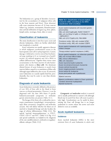
Table 13.1 Classifi cation of acute myeloid
ized by the accumulation of malignant white cells
leukaemia (AML) according to the WHO
in the bone marrow and blood. Th ese abnormal
classifi cation 2008 (modifi ed).
cells cause symptoms because of: (i) bone marrow
failure (e.g. anaemia, neutropenia, thrombocytope- Acute myeloid leukaemia with recurrent genetic
nia); and (ii) infiltration of organs (e.g. liver, spleen, abnormalities
lymph nodes, meninges, brain, skin or testes). AML with t(8;21)(q22;q22); RUNX1 - RUNX1T1
AML with inv(16)(p13.1q22) or t(16;6)(p13.1;q22);
CBFB - MYH11
Classification of l eukaemia
AML with t(15;17)(q22;q12); PML - RARA
The main classifi cation is into four types: acute and Provisional entity: AML with mutated NPM1
chronic leukaemias, which are further subdivided Provisional entity: AML with mutated CEBPA
into lymphoid or myeloid.
Acute myeloid leukaemia with myelodysplasia -
Acute leukaemias are usually aggressive diseases
related changes
in which malignant transformation occurs in the
haemopoietic stem cell or early progenitors. Genetic Therapy - related myeloid neoplasms (t - AML)
damage is believed to involve several key biochemi- Acute myeloid leukaemia, not otherwise specifi ed
cal steps resulting in (i) an increased rate of prolif- AML with minimal differentiation
eration, (ii) reduced apoptosis and (iii) a block in AML without differentiation
cellular differentiation. Together these events cause AML with maturation
accumulation in the bone marrow of early haemo- Acute myelomonocytic leukaemia
poietic cells known as blast cells . Th e dominant Acute monoblastic/monocytic leukaemia
clinical feature of acute leukaemia is usually bone Acute erythroid leukaemia
marrow failure caused by accumulation of blast cells Acute megakaryoblastic leukaemia
although organ infiltration also occurs. If untreated, Acute basophilic leukaemia
acute leukaemias are usually rapidly fatal but, para- Acute panmyelosis with myelofi brosis
doxically, they may be easier to cure than chronic Myeloid sarcoma
leukaemias.
Myeloid proliferations related to Down syndrome
Transient abnormal myelopoiesis
Diagnosis of a cute l eukaemia Myeloid leukaemia
Acute leukaemia is normally defined as the presence
of over 20% of blast cells in the blood or bone
marrow at clinical presentation. However, it can be
diagnosed with less than 20% blasts if specifi c Cytogenetic and molecular analysis is essential
leukaemia - associated cytogenetic or molecular and is usually performed on marrow cells although
genetic abnormalities are present (Table 13.1 ). blood may be used if the blast cell count is particu-
The lineage of the blast cells is defined by micro- larly high. Cytochemistry can be useful in deter-
scopic examination (morphology), immunopheno- mining the blast cell lineage but is no longer
typic (flow cytometry), cytogenetic and molecular performed in centres where the newer and more
analysis. This will define whether the blasts are of definitive tests are available.
myeloid or lymphoid lineage and also localize the
stage of cellular differentiation (Table 13.2 ). Th e Acute m yeloid l eukaemia
+
typical ‘ myeloid immunophenotype ’ is CD13 ,
+
+
−
CD33 , CD117 and TdT (Table 13.2 ; Fig. 13.1 ) Incidence
and special antibodies are helpful in the diagnosis
of the rare undifferentiated, erythroid or megakary- Acute myeloid leukaemia (AML) is the most
oblastic subtypes (Table 13.2 ). common form of acute leukaemia in adults and